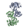
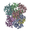
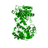
Journal: Proteins / Year: 2008 Title: Crystal Structures of the Mycobacterium Tuberculosis Secretory Antigen Alanine Dehydrogenase (Rv2780) in Apo and Ternary Complex Forms Captures "Open" and "Closed" Enzyme Conformations. Authors: Tripathi, S.M. / Ramachandran, R.
History
Deposition
Feb 17, 2008
Deposition site: PDBE / Processing site: PDBE
Revision 1.0
Mar 4, 2008
Provider: repository / Type: Initial release
Revision 1.1
Jan 18, 2012
Group: Database references / Derived calculations ...Database references / Derived calculations / Non-polymer description / Other / Refinement description / Structure summary / Version format compliance
Protocol: SINGLE WAVELENGTH / Monochromatic (M) / Laue (L): M / Scattering type: x-ray
Radiation wavelength
Wavelength: 1.5418 Å / Relative weight: 1
Reflection
Resolution: 2.6→25 Å / Num. obs: 46867 / % possible obs: 97.6 % / Observed criterion σ(I): 1 / Redundancy: 1.7 % / Rmerge(I) obs: 0.11 / Net I/σ(I): 9.6
-
Processing
Software
Name
Version
Classification
REFMAC
5.2.0005
refinement
HKL-2000
datareduction
SCALEPACK
datascaling
Refinement
Method to determine structure: OTHER Starting model: NONE Resolution: 2.6→123.09 Å / Cor.coef. Fo:Fc: 0.941 / Cor.coef. Fo:Fc free: 0.9 / SU B: 10.695 / SU ML: 0.226 / Cross valid method: THROUGHOUT / ESU R: 0.666 / ESU R Free: 0.313 / Stereochemistry target values: MAXIMUM LIKELIHOOD / Details: HYDROGENS HAVE BEEN ADDED IN THE RIDING POSITIONS.
Rfactor
Num. reflection
% reflection
Selection details
Rfree
0.2512
4005
5 %
RANDOM
Rwork
0.19561
-
-
-
obs
0.19837
75667
97.24 %
-
Solvent computation
Ion probe radii: 0.8 Å / Shrinkage radii: 0.8 Å / VDW probe radii: 1.2 Å / Solvent model: MASK
 Movie
Movie Controller


 Open data
 Basic information
 Components
 Keywords
 Function and homology information
 MYCOBACTERIUM TUBERCULOSIS (bacteria)
 X-RAY DIFFRACTION / OTHER / Resolution: 2.6 Å
  Authors
 Citation
 Structure visualization
 Downloads & links
 Other downloads
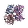


 PDBj
PDBj Assembly
 Mass: 18.015 Da / Num. of mol.: 224 / Source method: isolated from a natural source / Formula: H2O
 Sample preparation
 Processing