 Movie
Movie Controller

[English] 日本語
 Yorodumi
- PDB-4lmp: Mycobacterium tuberculosis L-alanine dehydrogenase x-ray structur... -
+ Open data

- Basic information
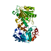
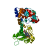
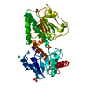
| Entry | Database: PDB / ID: 4lmp | ||||||
|---|---|---|---|---|---|---|---|
| Title | Mycobacterium tuberculosis L-alanine dehydrogenase x-ray structure in complex with N6-methyl adenosine | ||||||
 Components Components | Alanine dehydrogenase | ||||||
 Keywords Keywords | OXIDOREDUCTASE / Structural Genomics / PSI-Biology / Susceptibility to Known Mtb Inhibitors / MTBI / Rossmann Fold / N6-methyl adenosine binding / Structures of Mtb Proteins Conferring Susceptibility to Known Mtb Inhibitors | ||||||
| Function / homology | NAD(P)-binding Rossmann-like Domain / Rossmann fold / 3-Layer(aba) Sandwich / Alpha Beta / N-methyladenosine / :  Function and homology information Function and homology information | ||||||
| Biological species |   Mycobacterium tuberculosis (bacteria) Mycobacterium tuberculosis (bacteria) | ||||||
| Method |  X-RAY DIFFRACTION / SYNCHROTRON / MOLECULAR REPLACEMENT / Resolution: 1.95 Å X-RAY DIFFRACTION / SYNCHROTRON / MOLECULAR REPLACEMENT / Resolution: 1.95 Å | ||||||
 Authors Authors | Kim, H.-B. / Hung, L.-W. / Goulding, C.W. / Terwilliger, T.C. / Kim, C.-Y. / Structures of Mtb Proteins Conferring Susceptibility to Known Mtb Inhibitors (MTBI) | ||||||
 Citation Citation | Journal: To be Published Title: Drug target analysis by dye-ligand affinity chromatography Authors: Kim, H.-B. / Hung, L.-W. / Goulding, C.W. / Terwilliger, T.C. / Kim, C.-Y. | ||||||
| History |
|
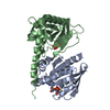
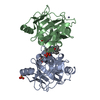
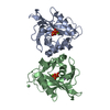
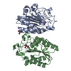
- Structure visualization
| Structure viewer | Molecule: MolmilJmol/JSmol |
|---|
- Downloads & links
-Download
| PDBx/mmCIF format | 4lmp.cif.gz | 89 KB | Display | PDBx/mmCIF format |
|---|---|---|---|---|
| PDB format | pdb4lmp.ent.gz | 66.3 KB | Display | PDB format |
| PDBx/mmJSON format | 4lmp.json.gz | Tree view | PDBx/mmJSON format | |
| Others |  Other downloads Other downloads |
-Validation report
| Arichive directory | https://data.pdbj.org/pub/pdb/validation_reports/lm/4lmpftp://data.pdbj.org/pub/pdb/validation_reports/lm/4lmp | HTTPS FTP |
|---|
-Related structure data
| Related structure data | 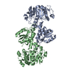 2vhyS S: Starting model for refinement |
|---|---|
| Similar structure data |
-Links
 PDBj
PDBj- Assembly
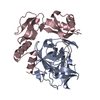
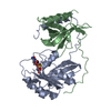
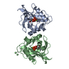
| Deposited unit | 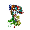
| ||||||||
|---|---|---|---|---|---|---|---|---|---|
| 1 |
| ||||||||
| 2 | 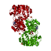
| ||||||||
| 3 | x 6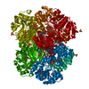
| ||||||||
| Unit cell |
| ||||||||
| Components on special symmetry positions |
|
-Components
| #1: Protein | Mass: 38753.090 Da / Num. of mol.: 1 Source method: isolated from a genetically manipulated source Source: (gene. exp.) Mycobacterium tuberculosis (bacteria) / Strain: H37Rv / Gene: ald, RVBD_2780 / Plasmid: pETM11 / Production host: | ||||
|---|---|---|---|---|---|
| #2: Chemical | ChemComp-6MD /   Mass: 281.268 Da / Num. of mol.: 1 / Source method: obtained synthetically / Formula: C11H15N5O4 Mass: 281.268 Da / Num. of mol.: 1 / Source method: obtained synthetically / Formula: C11H15N5O4 | ||||
| #3: Chemical | ChemComp-GOL /   Mass: 92.094 Da / Num. of mol.: 4 / Source method: obtained synthetically / Formula: C3H8O3 Mass: 92.094 Da / Num. of mol.: 4 / Source method: obtained synthetically / Formula: C3H8O3#4: Chemical |   Mass: 96.063 Da / Num. of mol.: 3 / Source method: obtained synthetically / Formula: SO4 Mass: 96.063 Da / Num. of mol.: 3 / Source method: obtained synthetically / Formula: SO4#5: Water | ChemComp-HOH / |  Mass: 18.015 Da / Num. of mol.: 211 / Source method: isolated from a natural source / Formula: H2O Mass: 18.015 Da / Num. of mol.: 211 / Source method: isolated from a natural source / Formula: H2O |
-Experimental details
-Experiment
| Experiment | Method: X-RAY DIFFRACTION / Number of used crystals: 1 |
|---|
- Sample preparation
| Crystal | Density Matthews: 2.9 Å3/Da / Density % sol: 57.63 % |
|---|---|
| Crystal grow | Temperature: 298 K / Method: vapor diffusion, hanging drop / pH: 5.4 Details: 2M ammonium sulfate, 5% 2-propanol, pH 5.4, VAPOR DIFFUSION, HANGING DROP, temperature 298.0K |
-Data collection
| Diffraction | Mean temperature: 100 K |
|---|---|
| Diffraction source | Source: SYNCHROTRON / Site: ALS  / Beamline: 5.0.1 / Wavelength: 0.9774 Å / Beamline: 5.0.1 / Wavelength: 0.9774 Å |
| Detector | Type: ADSC QUANTUM 315r / Detector: CCD / Date: Jan 22, 2010 / Details: Si111 |
| Radiation | Monochromator: Si111 / Protocol: SINGLE WAVELENGTH / Monochromatic (M) / Laue (L): M / Scattering type: x-ray |
| Radiation wavelength | Wavelength: 0.9774 Å / Relative weight: 1 |
| Reflection | Resolution: 1.95→50 Å / Num. all: 33325 / Num. obs: 33325 / % possible obs: 100 % / Observed criterion σ(F): 0 / Observed criterion σ(I): -3 / Redundancy: 5.8 % / Biso Wilson estimate: 23.97 Å2 / Rmerge(I) obs: 0.072 / Net I/σ(I): 22.84 |
| Reflection shell | Resolution: 1.95→1.98 Å / Redundancy: 4.1 % / Rmerge(I) obs: 0.478 / Mean I/σ(I) obs: 2.234 / Num. unique all: 1602 / % possible all: 99 |
- Processing
| Software |
| |||||||||||||||||||||||||||||||||||||||||||||||||||||||||||||||||||||||||||||||||||||||||||||||||||||||||
|---|---|---|---|---|---|---|---|---|---|---|---|---|---|---|---|---|---|---|---|---|---|---|---|---|---|---|---|---|---|---|---|---|---|---|---|---|---|---|---|---|---|---|---|---|---|---|---|---|---|---|---|---|---|---|---|---|---|---|---|---|---|---|---|---|---|---|---|---|---|---|---|---|---|---|---|---|---|---|---|---|---|---|---|---|---|---|---|---|---|---|---|---|---|---|---|---|---|---|---|---|---|---|---|---|---|---|
| Refinement | Method to determine structure: MOLECULAR REPLACEMENT Starting model: PDB ENTRY 2VHY Resolution: 1.95→48.4 Å / SU ML: 0.2 / Cross valid method: THROUGHOUT / σ(F): 0 / Phase error: 21.68 / Stereochemistry target values: ML
| |||||||||||||||||||||||||||||||||||||||||||||||||||||||||||||||||||||||||||||||||||||||||||||||||||||||||
| Solvent computation | Shrinkage radii: 0.9 Å / VDW probe radii: 1.11 Å / Solvent model: FLAT BULK SOLVENT MODEL | |||||||||||||||||||||||||||||||||||||||||||||||||||||||||||||||||||||||||||||||||||||||||||||||||||||||||
| Displacement parameters | Biso mean: 23.97 Å2 | |||||||||||||||||||||||||||||||||||||||||||||||||||||||||||||||||||||||||||||||||||||||||||||||||||||||||
| Refinement step | Cycle: LAST / Resolution: 1.95→48.4 Å
| |||||||||||||||||||||||||||||||||||||||||||||||||||||||||||||||||||||||||||||||||||||||||||||||||||||||||
| Refine LS restraints |
| |||||||||||||||||||||||||||||||||||||||||||||||||||||||||||||||||||||||||||||||||||||||||||||||||||||||||
| LS refinement shell |
|
