Protocol: SINGLE WAVELENGTH / Monochromatic (M) / Laue (L): M / Scattering type: x-ray
Radiation wavelength
Wavelength: 0.979 Å / Relative weight: 1
Reflection
Resolution: 1.29→50 Å / Num. obs: 115974 / % possible obs: 99.2 % / Redundancy: 12.3 % / Net I/σ(I): 34.9
-
Processing
Software
Name
Version
Classification
REFMAC
5.8.0073
refinement
HKL-3000
datascaling
HKL-3000
phasing
Refinement
Resolution: 1.68→50 Å / Cor.coef. Fo:Fc: 0.965 / Cor.coef. Fo:Fc free: 0.928 / SU B: 1.445 / SU ML: 0.05 / Cross valid method: THROUGHOUT / ESU R: 0.085 / ESU R Free: 0.088 / Details: HYDROGENS HAVE BEEN ADDED IN THE RIDING POSITIONS
Rfactor
Num. reflection
% reflection
Selection details
Rfree
0.18715
2827
5 %
RANDOM
Rwork
0.15186
-
-
-
obs
0.15364
53665
99.74 %
-
Solvent computation
Ion probe radii: 0.8 Å / Shrinkage radii: 0.8 Å / VDW probe radii: 1.2 Å
 Movie
Movie Controller

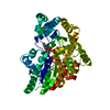
 Yorodumi
 Open data
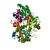
 Basic information
 Components
 Keywords
 Function and homology information
 X-RAY DIFFRACTION /
  Authors
 Citation
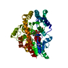
 Structure visualization
 Downloads & links
 Other downloads
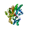


 PDBj
PDBj


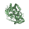
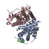
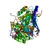
 Assembly


 Type: RNA linking / Mass: 404.161 Da / Num. of mol.: 1 / Source method: obtained synthetically / Formula: C9H14N2O12P2 / Comment: UDP*YM
 Mass: 18.015 Da / Num. of mol.: 486 / Source method: isolated from a natural source / Formula: H2O
 Sample preparation
 / Beamline: 19-ID / Wavelength: 0.979 Å
 Processing