 Movie
Movie Controller

[English] 日本語
 Yorodumi
- PDB-5m8m: Crystal structure of human tyrosinase related protein 1 in comple... -
+ Open data

- Basic information
| Entry | Database: PDB / ID: 5m8m | ||||||||||||
|---|---|---|---|---|---|---|---|---|---|---|---|---|---|
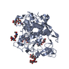
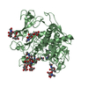
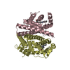
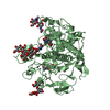
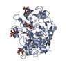
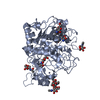
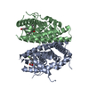
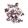
| Title | Crystal structure of human tyrosinase related protein 1 in complex with kojic acid | ||||||||||||
 Components Components | 5,6-dihydroxyindole-2-carboxylic acid oxidase | ||||||||||||
 Keywords Keywords | UNKNOWN FUNCTION / human tyrosinase related protein 1 / melanin biosynthesis / tyrosinase / oxidoreductase | ||||||||||||
| Function / homology |  Function and homology information Function and homology information: / Melanin biosynthesis / Oxidoreductases; Acting on paired donors, with incorporation or reduction of molecular oxygen; With another compound as one donor, and incorporation of one atom of oxygen into the other donor / tyrosinase activity / catechol oxidase activity / positive regulation of melanin biosynthetic process / melanin biosynthetic process / melanocyte differentiation / melanosome membrane / melanosome organization ...: / Melanin biosynthesis / Oxidoreductases; Acting on paired donors, with incorporation or reduction of molecular oxygen; With another compound as one donor, and incorporation of one atom of oxygen into the other donor / tyrosinase activity / catechol oxidase activity / positive regulation of melanin biosynthetic process / melanin biosynthetic process / melanocyte differentiation / melanosome membrane / melanosome organization / intracellular vesicle / Regulation of MITF-M-dependent genes involved in pigmentation / clathrin-coated endocytic vesicle membrane / melanosome / endosome membrane / protein homodimerization activity / metal ion binding / cytoplasm Similarity search - Function | ||||||||||||
| Biological species |  Homo sapiens (human) Homo sapiens (human) | ||||||||||||
| Method |  X-RAY DIFFRACTION / SYNCHROTRON / MOLECULAR REPLACEMENT / Resolution: 2.65 Å X-RAY DIFFRACTION / SYNCHROTRON / MOLECULAR REPLACEMENT / Resolution: 2.65 Å | ||||||||||||
 Authors Authors | Lai, X. / Soler-Lopez, M. / Wichers, H.J. / Dijkstra, B.W. | ||||||||||||
 Citation Citation | Journal: Angew. Chem. Int. Ed. Engl. / Year: 2017 Title: Structure of Human Tyrosinase Related Protein 1 Reveals a Binuclear Zinc Active Site Important for Melanogenesis. Authors: Lai, X. / Wichers, H.J. / Soler-Lopez, M. / Dijkstra, B.W. | ||||||||||||
| History |
|
- Structure visualization
| Structure viewer | Molecule: MolmilJmol/JSmol |
|---|
- Downloads & links
-Download
| PDBx/mmCIF format | 5m8m.cif.gz | 374 KB | Display | PDBx/mmCIF format |
|---|---|---|---|---|
| PDB format | pdb5m8m.ent.gz | 307.1 KB | Display | PDB format |
| PDBx/mmJSON format | 5m8m.json.gz | Tree view | PDBx/mmJSON format | |
| Others |  Other downloads Other downloads |
-Validation report
| Arichive directory | https://data.pdbj.org/pub/pdb/validation_reports/m8/5m8mftp://data.pdbj.org/pub/pdb/validation_reports/m8/5m8m | HTTPS FTP |
|---|
-Related structure data
| Related structure data |  5m8lC  5m8nC  5m8oC  5m8pC  5m8qC  5m8rC  5m8tC C: citing same article ( |
|---|---|
| Similar structure data |
-Links
 PDBj
PDBj- Assembly
| Deposited unit | 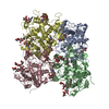
| ||||||||
|---|---|---|---|---|---|---|---|---|---|
| 1 | 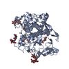
| ||||||||
| 2 | 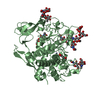
| ||||||||
| 3 | 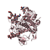
| ||||||||
| 4 | 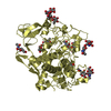
| ||||||||
| Unit cell |
|
-Components
-Protein , 1 types, 4 molecules ABCD
| #1: Protein | Mass: 50619.250 Da / Num. of mol.: 4 Source method: isolated from a genetically manipulated source Source: (gene. exp.) Homo sapiens (human) / Gene: TYRP1, CAS2, TYRP, TYRRP / Production host:   Spodoptera frugiperda (fall armyworm) Spodoptera frugiperda (fall armyworm)References: UniProt: P17643, Oxidoreductases; Acting on paired donors, with incorporation or reduction of molecular oxygen; With another compound as one donor, and incorporation of one atom of oxygen into the other donor |
|---|
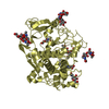
-Sugars , 7 types, 21 molecules 
| #2: Polysaccharide | 2-acetamido-2-deoxy-beta-D-glucopyranose-(1-4)-[alpha-L-fucopyranose-(1-6)]2-acetamido-2-deoxy-beta- ...2-acetamido-2-deoxy-beta-D-glucopyranose-(1-4)-[alpha-L-fucopyranose-(1-6)]2-acetamido-2-deoxy-beta-D-glucopyranose Source method: isolated from a genetically manipulated source #3: Polysaccharide | Source method: isolated from a genetically manipulated source #4: Polysaccharide | 2-acetamido-2-deoxy-beta-D-glucopyranose-(1-4)-2-acetamido-2-deoxy-beta-D-glucopyranose Source method: isolated from a genetically manipulated source #5: Polysaccharide | alpha-D-mannopyranose-(1-3)-alpha-D-mannopyranose-(1-4)-2-acetamido-2-deoxy-beta-D-glucopyranose-(1- ...alpha-D-mannopyranose-(1-3)-alpha-D-mannopyranose-(1-4)-2-acetamido-2-deoxy-beta-D-glucopyranose-(1-4)-2-acetamido-2-deoxy-beta-D-glucopyranose | Source method: isolated from a genetically manipulated source #6: Polysaccharide | alpha-D-mannopyranose-(1-3)-alpha-D-mannopyranose-(1-4)-2-acetamido-2-deoxy-beta-D-glucopyranose-(1- ...alpha-D-mannopyranose-(1-3)-alpha-D-mannopyranose-(1-4)-2-acetamido-2-deoxy-beta-D-glucopyranose-(1-4)-[alpha-L-fucopyranose-(1-6)]2-acetamido-2-deoxy-beta-D-glucopyranose | Source method: isolated from a genetically manipulated source #7: Polysaccharide | alpha-D-mannopyranose-(1-3)-[alpha-D-mannopyranose-(1-6)]alpha-D-mannopyranose-(1-4)-2-acetamido-2- ...alpha-D-mannopyranose-(1-3)-[alpha-D-mannopyranose-(1-6)]alpha-D-mannopyranose-(1-4)-2-acetamido-2-deoxy-beta-D-glucopyranose-(1-4)-2-acetamido-2-deoxy-beta-D-glucopyranose | Source method: isolated from a genetically manipulated source #10: Sugar | ChemComp-NAG / |  Type: D-saccharide, beta linking / Mass: 221.208 Da / Num. of mol.: 1 Type: D-saccharide, beta linking / Mass: 221.208 Da / Num. of mol.: 1Source method: isolated from a genetically manipulated source Formula: C8H15NO6 |
|---|
-Non-polymers , 3 types, 56 molecules 
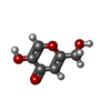

| #8: Chemical | ChemComp-ZN /  Mass: 65.409 Da / Num. of mol.: 9 / Source method: obtained synthetically / Formula: Zn Mass: 65.409 Da / Num. of mol.: 9 / Source method: obtained synthetically / Formula: Zn#9: Chemical | ChemComp-KOJ /  Mass: 142.109 Da / Num. of mol.: 4 / Source method: obtained synthetically / Formula: C6H6O4 Mass: 142.109 Da / Num. of mol.: 4 / Source method: obtained synthetically / Formula: C6H6O4#11: Water | ChemComp-HOH / | Mass: 18.015 Da / Num. of mol.: 43 / Source method: isolated from a natural source / Formula: H2O |
|---|
-Details
| Has protein modification | Y |
|---|
-Experimental details
-Experiment
| Experiment | Method: X-RAY DIFFRACTION / Number of used crystals: 1 |
|---|
- Sample preparation
| Crystal | Density Matthews: 3.03 Å3/Da / Density % sol: 59.41 % |
|---|---|
| Crystal grow | Temperature: 290 K / Method: vapor diffusion, sitting drop Details: 0.1 M Tris (pH 7.0), 0.2 M NaCl and 30% (w/v) PEG 3000 |
-Data collection
| Diffraction | Mean temperature: 100 K |
|---|---|
| Diffraction source | Source: SYNCHROTRON / Site: ESRF  / Beamline: ID29 / Wavelength: 0.976 Å / Beamline: ID29 / Wavelength: 0.976 Å |
| Detector | Type: DECTRIS PILATUS 6M-F / Detector: PIXEL / Date: Sep 1, 2015 |
| Radiation | Protocol: SINGLE WAVELENGTH / Monochromatic (M) / Laue (L): M / Scattering type: x-ray |
| Radiation wavelength | Wavelength: 0.976 Å / Relative weight: 1 |
| Reflection | Resolution: 2.65→49 Å / Num. obs: 71959 / % possible obs: 99.8 % / Redundancy: 4.6 % / Net I/σ(I): 9 |
- Processing
| Software |
| ||||||||||||||||||||||||
|---|---|---|---|---|---|---|---|---|---|---|---|---|---|---|---|---|---|---|---|---|---|---|---|---|---|
| Refinement | Method to determine structure: MOLECULAR REPLACEMENT / Resolution: 2.65→49 Å / SU ML: 0.37 / Cross valid method: FREE R-VALUE / σ(F): 1.34 / Phase error: 25.64
| ||||||||||||||||||||||||
| Solvent computation | Shrinkage radii: 0.9 Å / VDW probe radii: 1.11 Å | ||||||||||||||||||||||||
| Displacement parameters | Biso max: 138.16 Å2 / Biso mean: 39.6045 Å2 / Biso min: 15.12 Å2 | ||||||||||||||||||||||||
| Refinement step | Cycle: final / Resolution: 2.65→49 Å
|
