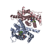
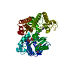
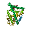
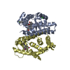
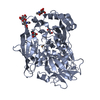
Entry Database : PDB / ID : 1xqcTitle X-ray structure of ERalpha LBD bound to a tetrahydroisoquinoline SERM ligand at 2.05A resolution Estrogen receptor Keywords / / / Function / homology Function Domain/homology Component
/ / / / / / / / / / / / / / / / / / / / / / / / / / / / / / / / / / / / / / / / / / / / / / / / / / / / / / / / / / / / / / / / / / / / / / / / / / / / / / / / / / / / / / / / / / / / / / / / / / / / / / / / / / / / / / / / / / / / / / / / / / / / / / / / / / / / / / Biological species Homo sapiens (human)Method / / / Resolution : 2.05 Å Authors Renaud, J. / Bischoff, S.F. / Buhl, T. / Floersheim, P. / Fournier, B. / Geiser, M. / Halleux, C. / Kallen, J. / Keller, H.J. / Ramage, P. Journal : J.Med.Chem. / Year : 2005Title : Selective Estrogen Receptor Modulators with Conformationally Restricted Side Chains. Synthesis and Structure-Activity Relationship of ERalpha-Selective Tetrahydroisoquinoline LigandsAuthors : Renaud, J. / Bischoff, S.F. / Buhl, T. / Floersheim, P. / Fournier, B. / Geiser, M. / Halleux, C. / Kallen, J. / Keller, H.J. / Ramage, P. History Deposition Oct 12, 2004 Deposition site / Processing site Revision 1.0 Feb 1, 2005 Provider / Type Revision 1.1 Apr 30, 2008 Group Revision 1.2 Jul 13, 2011 Group Revision 1.3 Nov 10, 2021 Group / Derived calculations / Category / struct_ref_seq_dif / struct_siteItem _database_2.pdbx_DOI / _database_2.pdbx_database_accession ... _database_2.pdbx_DOI / _database_2.pdbx_database_accession / _struct_ref_seq_dif.details / _struct_site.pdbx_auth_asym_id / _struct_site.pdbx_auth_comp_id / _struct_site.pdbx_auth_seq_id Revision 1.4 Oct 25, 2023 Group / Refinement descriptionCategory / chem_comp_bond / pdbx_initial_refinement_model
Show all Show less
 Movie
Movie Controller

 Yorodumi
 Open data
 Basic information
 Components
 Keywords
 Function and homology information
 Homo sapiens (human)
 X-RAY DIFFRACTION /
  Authors
 Citation
 Structure visualization
 Downloads & links
 Other downloads


 PDBj
PDBj


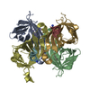
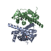
 Assembly
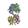


 Mass: 439.592 Da / Num. of mol.: 4 / Source method: obtained synthetically / Formula: C29H33N3O
 Mass: 18.015 Da / Num. of mol.: 374 / Source method: isolated from a natural source / Formula: H2O
 Sample preparation
 Processing