 Movie
Movie Controller


+ Open data

- Basic information
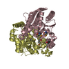
| Entry | Database: PDB / ID: 6lf6 | ||||||
|---|---|---|---|---|---|---|---|
| Title | Crystal structure of ZmCGTa in complex with UDP | ||||||
 Components Components | UDP-glycosyltransferase 708A6 | ||||||
 Keywords Keywords | TRANSFERASE / ZmCGTa / UDP / Complex | ||||||
| Function / homology |  Function and homology information Function and homology informationflavanone 7-O-beta-glucosyltransferase activity / UDP-glucosyltransferase activity / Transferases; Glycosyltransferases; Hexosyltransferases Similarity search - Function | ||||||
| Biological species |  | ||||||
| Method |  X-RAY DIFFRACTION / SYNCHROTRON / MOLECULAR REPLACEMENT / Resolution: 2.044 Å X-RAY DIFFRACTION / SYNCHROTRON / MOLECULAR REPLACEMENT / Resolution: 2.044 Å | ||||||
 Authors Authors | Gao, H.M. / Yun, C.H. | ||||||
 Citation Citation | Journal: Proc.Natl.Acad.Sci.USA / Year: 2020 Title: Dissection of the general two-step di- C -glycosylation pathway for the biosynthesis of (iso)schaftosides in higher plants. Authors: Wang, Z.L. / Gao, H.M. / Wang, S. / Zhang, M. / Chen, K. / Zhang, Y.Q. / Wang, H.D. / Han, B.Y. / Xu, L.L. / Song, T.Q. / Yun, C.H. / Qiao, X. / Ye, M. | ||||||
| History |
|
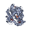
- Structure visualization
| Structure viewer | Molecule: MolmilJmol/JSmol |
|---|
- Downloads & links
-Download
| PDBx/mmCIF format | 6lf6.cif.gz | 104.2 KB | Display | PDBx/mmCIF format |
|---|---|---|---|---|
| PDB format | pdb6lf6.ent.gz | 75.9 KB | Display | PDB format |
| PDBx/mmJSON format | 6lf6.json.gz | Tree view | PDBx/mmJSON format | |
| Others |  Other downloads Other downloads |
-Validation report
| Arichive directory | https://data.pdbj.org/pub/pdb/validation_reports/lf/6lf6ftp://data.pdbj.org/pub/pdb/validation_reports/lf/6lf6 | HTTPS FTP |
|---|
-Related structure data
| Related structure data | 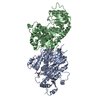 6lfnC 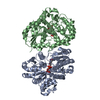 6lfzC  6lg0C  6lg1C 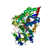 2vceS S: Starting model for refinement C: citing same article ( |
|---|---|
| Similar structure data |


-Links
 PDBj
PDBj


- Assembly
| Deposited unit | 
| ||||||||
|---|---|---|---|---|---|---|---|---|---|
| 1 |
| ||||||||
| Unit cell |
|
-Components
| #1: Protein | Mass: 50711.633 Da / Num. of mol.: 1 Source method: isolated from a genetically manipulated source Source: (gene. exp.)  References: UniProt: A0A096SRM5, Transferases; Glycosyltransferases; Hexosyltransferases |
|---|---|
| #2: Chemical | ChemComp-UDP /   Type: RNA linking / Mass: 404.161 Da / Num. of mol.: 1 / Source method: obtained synthetically / Formula: C9H14N2O12P2 / Comment: UDP*YM Type: RNA linking / Mass: 404.161 Da / Num. of mol.: 1 / Source method: obtained synthetically / Formula: C9H14N2O12P2 / Comment: UDP*YM |
| #3: Water | ChemComp-HOH /  Mass: 18.015 Da / Num. of mol.: 141 / Source method: isolated from a natural source / Formula: H2O Mass: 18.015 Da / Num. of mol.: 141 / Source method: isolated from a natural source / Formula: H2O |
| Has ligand of interest | Y |
-Experimental details
-Experiment
| Experiment | Method: X-RAY DIFFRACTION / Number of used crystals: 1 |
|---|
- Sample preparation
| Crystal | Density Matthews: 2.39 Å3/Da / Density % sol: 48.55 % |
|---|---|
| Crystal grow | Temperature: 298 K / Method: vapor diffusion, sitting drop / pH: 8.5 Details: 0.2M Trimethylamine N-oxide dihydrate, 0.1M Tris pH 8.5, 20% w/v Polyethylene glycol monomethyl ether 2000 |
-Data collection
| Diffraction | Mean temperature: 100 K / Serial crystal experiment: N |
|---|---|
| Diffraction source | Source: SYNCHROTRON / Site: SSRF  / Beamline: BL18U1 / Wavelength: 0.97853 Å / Beamline: BL18U1 / Wavelength: 0.97853 Å |
| Detector | Type: DECTRIS PILATUS3 6M / Detector: PIXEL / Date: May 24, 2019 |
| Radiation | Protocol: SINGLE WAVELENGTH / Monochromatic (M) / Laue (L): M / Scattering type: x-ray |
| Radiation wavelength | Wavelength: 0.97853 Å / Relative weight: 1 |
| Reflection | Resolution: 2.044→50 Å / Num. obs: 29864 / % possible obs: 99 % / Redundancy: 3.2 % / Biso Wilson estimate: 25.77 Å2 / Rpim(I) all: 0.04 / Net I/σ(I): 17.6 |
| Reflection shell | Resolution: 2.05→2.09 Å / Num. unique obs: 28126 / Rpim(I) all: 0.297 |
- Processing
| Software |
| ||||||||||||||||||||||||||||||||||||||||||||||||||||||||||||||||||
|---|---|---|---|---|---|---|---|---|---|---|---|---|---|---|---|---|---|---|---|---|---|---|---|---|---|---|---|---|---|---|---|---|---|---|---|---|---|---|---|---|---|---|---|---|---|---|---|---|---|---|---|---|---|---|---|---|---|---|---|---|---|---|---|---|---|---|---|
| Refinement | Method to determine structure: MOLECULAR REPLACEMENT Starting model: 2VCE Resolution: 2.044→28.034 Å / SU ML: 0.18 / Cross valid method: THROUGHOUT / σ(F): 1.37 / Phase error: 21.02 / Stereochemistry target values: ML
| ||||||||||||||||||||||||||||||||||||||||||||||||||||||||||||||||||
| Solvent computation | Shrinkage radii: 0.9 Å / VDW probe radii: 1.11 Å / Solvent model: FLAT BULK SOLVENT MODEL | ||||||||||||||||||||||||||||||||||||||||||||||||||||||||||||||||||
| Displacement parameters | Biso max: 75.15 Å2 / Biso mean: 27.2757 Å2 / Biso min: 12.14 Å2 | ||||||||||||||||||||||||||||||||||||||||||||||||||||||||||||||||||
| Refinement step | Cycle: final / Resolution: 2.044→28.034 Å
| ||||||||||||||||||||||||||||||||||||||||||||||||||||||||||||||||||
| LS refinement shell | Refine-ID: X-RAY DIFFRACTION / Rfactor Rfree error: 0
|
