 Movie
Movie Controller

[English] 日本語
 Yorodumi
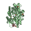
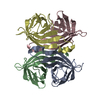
- PDB-3d24: Crystal structure of ligand-binding domain of estrogen-related re... -
+ Open data

- Basic information
| Entry | Database: PDB / ID: 3d24 | ||||||
|---|---|---|---|---|---|---|---|
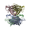
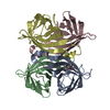
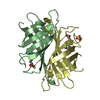
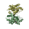
| Title | Crystal structure of ligand-binding domain of estrogen-related receptor alpha (ERRalpha) in complex with the peroxisome proliferators-activated receptor coactivator-1alpha box3 peptide (PGC-1alpha) | ||||||
 Components Components |
| ||||||
 Keywords Keywords | TRANSCRIPTION / nuclear receptor / ligand binding domain / coactivator / DNA-binding / Metal-binding / Nucleus / Phosphoprotein / Transcription regulation / Zinc / Zinc-finger / Polymorphism / RNA-binding / Structural Genomics / Structural Proteomics in Europe / SPINE | ||||||
| Function / homology |  Function and homology information Function and homology informationRegulation of MITF-M dependent genes involved in metabolism / positive regulation of fatty acid oxidation / Activation of PPARGC1A (PGC-1alpha) by phosphorylation / cellular respiration / response to muscle activity / response to starvation / nuclear steroid receptor activity / response to dietary excess / lncRNA binding / fatty acid oxidation ...Regulation of MITF-M dependent genes involved in metabolism / positive regulation of fatty acid oxidation / Activation of PPARGC1A (PGC-1alpha) by phosphorylation / cellular respiration / response to muscle activity / response to starvation / nuclear steroid receptor activity / response to dietary excess / lncRNA binding / fatty acid oxidation / temperature homeostasis / adipose tissue development / estrogen response element binding / FOXO-mediated transcription of oxidative stress, metabolic and neuronal genes / energy homeostasis / Transcriptional regulation of brown and beige adipocyte differentiation by EBF2 / digestion / intercellular bridge / steroid binding / RORA,B,C and NR1D1 (REV-ERBA) regulate gene expression / SUMOylation of transcription cofactors / Expression of BMAL (ARNTL), CLOCK, and NPAS2 / positive regulation of gluconeogenesis / brown fat cell differentiation / intracellular glucose homeostasis / RNA splicing / nuclear receptor binding / gluconeogenesis / respiratory electron transport chain / circadian regulation of gene expression / transcription coregulator activity / negative regulation of smooth muscle cell proliferation / mitochondrion organization / Heme signaling / transcription initiation at RNA polymerase II promoter / PPARA activates gene expression / Transcriptional activation of mitochondrial biogenesis / Nuclear Receptor transcription pathway / Transcriptional regulation of white adipocyte differentiation / chromatin DNA binding / regulation of circadian rhythm / PML body / DNA-binding transcription repressor activity, RNA polymerase II-specific / fibrillar center / nuclear receptor activity / mRNA processing / sequence-specific double-stranded DNA binding / Regulation of RUNX2 expression and activity / neuron apoptotic process / positive regulation of cold-induced thermogenesis / microtubule cytoskeleton / MLL4 and MLL3 complexes regulate expression of PPARG target genes in adipogenesis and hepatic steatosis / cellular response to oxidative stress / protein-containing complex assembly / DNA-binding transcription activator activity, RNA polymerase II-specific / DNA-binding transcription factor binding / sequence-specific DNA binding / negative regulation of neuron apoptotic process / RNA polymerase II-specific DNA-binding transcription factor binding / DNA-binding transcription factor activity, RNA polymerase II-specific / transcription coactivator activity / protein stabilization / DNA-binding transcription factor activity / protein domain specific binding / positive regulation of gene expression / ubiquitin protein ligase binding / regulation of transcription by RNA polymerase II / regulation of DNA-templated transcription / positive regulation of DNA-templated transcription / chromatin / positive regulation of transcription by RNA polymerase II / DNA binding / RNA binding / zinc ion binding / nucleoplasm / nucleus / cytosol / cytoplasm Similarity search - Function | ||||||
| Biological species |  Homo sapiens (human) Homo sapiens (human) | ||||||
| Method |  X-RAY DIFFRACTION / SYNCHROTRON / MOLECULAR REPLACEMENT / Resolution: 2.11 Å X-RAY DIFFRACTION / SYNCHROTRON / MOLECULAR REPLACEMENT / Resolution: 2.11 Å | ||||||
 Authors Authors | Moras, D. / Greschik, H. / Flaig, R. / Sato, Y. / Rochel, N. / Structural Proteomics in Europe (SPINE) | ||||||
 Citation Citation | Journal: J.Biol.Chem. / Year: 2008 Title: Communication between the ERR{alpha} Homodimer Interface and the PGC-1{alpha} Binding Surface via the Helix 8-9 Loop. Authors: Greschik, H. / Althage, M. / Flaig, R. / Sato, Y. / Chavant, V. / Peluso-Iltis, C. / Choulier, L. / Cronet, P. / Rochel, N. / Schule, R. / Stromstedt, P.E. / Moras, D. | ||||||
| History |
|
- Structure visualization
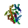
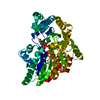
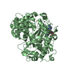
| Structure viewer | Molecule: MolmilJmol/JSmol |
|---|
- Downloads & links
-Download
| PDBx/mmCIF format | 3d24.cif.gz | 106.9 KB | Display | PDBx/mmCIF format |
|---|---|---|---|---|
| PDB format | pdb3d24.ent.gz | 81.8 KB | Display | PDB format |
| PDBx/mmJSON format | 3d24.json.gz | Tree view | PDBx/mmJSON format | |
| Others |  Other downloads Other downloads |
-Validation report
| Arichive directory | https://data.pdbj.org/pub/pdb/validation_reports/d2/3d24ftp://data.pdbj.org/pub/pdb/validation_reports/d2/3d24 | HTTPS FTP |
|---|
-Related structure data
| Related structure data |  1xb7S S: Starting model for refinement |
|---|---|
| Similar structure data | |
| Other databases |
-Links
 PDBj
PDBj


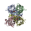
- Assembly
| Deposited unit | 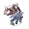
| ||||||||
|---|---|---|---|---|---|---|---|---|---|
| 1 |
| ||||||||
| 2 | 
| ||||||||
| 3 | 
| ||||||||
| Unit cell |
| ||||||||
| Details | AUTHORS STATE THAT THE BIOLOGICAL ASSEMBLY IS A HOMODIMER. |
-Components
| #1: Protein | Mass: 27584.893 Da / Num. of mol.: 2 / Fragment: Ligand binding domain: Residues 278-519 Source method: isolated from a genetically manipulated source Source: (gene. exp.) Homo sapiens (human) / Gene: ESRRA, ERR1, ESRL1, NR3B1 / Plasmid: pET24 / Production host:  #2: Protein/peptide | Mass: 2665.975 Da / Num. of mol.: 2 / Fragment: Residues 198-219 / Source method: obtained synthetically Details: Synthetic peptide with the sequence based on the fragment (residues 198-219) of human PGC-1-alpha, UniProt entry Q9UBK2 (PRGC1_HUMAN) References: UniProt: Q9UBK2 #3: Water | ChemComp-HOH / |  Mass: 18.015 Da / Num. of mol.: 272 / Source method: isolated from a natural source / Formula: H2O Mass: 18.015 Da / Num. of mol.: 272 / Source method: isolated from a natural source / Formula: H2O |
|---|
-Experimental details
-Experiment
| Experiment | Method: X-RAY DIFFRACTION / Number of used crystals: 1 |
|---|
- Sample preparation
| Crystal | Density Matthews: 2.47 Å3/Da / Density % sol: 50.28 % |
|---|---|
| Crystal grow | Temperature: 290 K / Method: vapor diffusion, hanging drop / pH: 5.5 Details: 0.1 M Bis-Tris pH 5.5, 15% PEG 3350, 0.2 M Mg(NO3)2, VAPOR DIFFUSION, HANGING DROP, temperature 290K |
-Data collection
| Diffraction | Mean temperature: 100 K |
|---|---|
| Diffraction source | Source: SYNCHROTRON / Site: ESRF  / Beamline: ID14-2 / Wavelength: 0.933 Å / Beamline: ID14-2 / Wavelength: 0.933 Å |
| Detector | Type: ADSC QUANTUM 4 / Detector: CCD / Date: Sep 13, 2007 / Details: Toroidal Zerodur mirror |
| Radiation | Monochromator: Si 111 CHANNEL / Protocol: SINGLE WAVELENGTH / Monochromatic (M) / Laue (L): M / Scattering type: x-ray |
| Radiation wavelength | Wavelength: 0.933 Å / Relative weight: 1 |
| Reflection | Resolution: 2.11→50 Å / Num. all: 111465 / Num. obs: 109542 / % possible obs: 88.9 % / Observed criterion σ(I): 2 / Redundancy: 3.5 % / Biso Wilson estimate: 29.7 Å2 / Rsym value: 0.031 / Net I/σ(I): 26.2 |
| Reflection shell | Resolution: 2.11→2.19 Å / Redundancy: 3.2 % / Num. unique all: 2740 / Rsym value: 0.238 / % possible all: 76.9 |
- Processing
| Software |
| |||||||||||||||||||||||||
|---|---|---|---|---|---|---|---|---|---|---|---|---|---|---|---|---|---|---|---|---|---|---|---|---|---|---|
| Refinement | Method to determine structure: MOLECULAR REPLACEMENT Starting model: PDB entry 1XB7 Resolution: 2.11→50 Å / σ(F): 0 / σ(I): 0 / Stereochemistry target values: MAXIMUM LIKELIHOOD / Details: The Bijvoet differences were used in phasing
| |||||||||||||||||||||||||
| Displacement parameters | Biso mean: 42.5 Å2
| |||||||||||||||||||||||||
| Refinement step | Cycle: LAST / Resolution: 2.11→50 Å
| |||||||||||||||||||||||||
| Refine LS restraints |
|
