Mass: 18.015 Da / Num. of mol.: 508 / Source method: isolated from a natural source / Formula: H2O
-
Details
Has protein modification
Y
Sequence details
THE CONSTRUCT WAS EXPRESSED WITH A PURIFICATION TAG MGSDKIHHHHHHENLYFQG. THE TAG WAS REMOVED WITH ...THE CONSTRUCT WAS EXPRESSED WITH A PURIFICATION TAG MGSDKIHHHHHHENLYFQG. THE TAG WAS REMOVED WITH TEV PROTEASE LEAVING ONLY A GLYCINE (0) FOLLOWED BY THE TARGET SEQUENCE.
-
Experimental details
-
Experiment
Experiment
Method: X-RAY DIFFRACTION / Number of used crystals: 1
-
Sample preparation
Crystal
Density Matthews: 3.16 Å3/Da / Density % sol: 61.09 %
Crystal grow
Temperature: 277 K / Method: vapor diffusion, sitting drop / pH: 4.5 Details: 40.0000% 1,2-propanediol, 0.0500M Ca(OAc)2, 0.1M Acetate pH 4.5, NANODROP, VAPOR DIFFUSION, SITTING DROP, temperature 277K
Monochromator: Single crystal Si(311) bent monochromator (horizontal focusing) Protocol: MAD / Monochromatic (M) / Laue (L): M / Scattering type: x-ray
Radiation wavelength
ID
Wavelength (Å)
Relative weight
1
0.91837
1
2
0.978985
1
3
0.979346
1
Reflection
Resolution: 1.8→30.056 Å / Num. obs: 68777 / % possible obs: 99.6 % / Observed criterion σ(I): -3 / Redundancy: 7.3 % / Biso Wilson estimate: 20.106 Å2 / Rmerge F obs: 0.126 / Rmerge(I) obs: 0.068 / Net I/σ(I): 14.56
Reflection shell
Resolution (Å)
Rmerge(I) obs
Mean I/σ(I) obs
Num. measured obs
Num. unique obs
Diffraction-ID
% possible all
1.8-1.86
0.618
2.3
45842
12088
1
97.9
1.86-1.94
0.438
3.3
54015
14207
1
99.6
1.94-2.03
0.29
5
51049
13416
1
99.7
2.03-2.13
0.202
7.1
47314
12428
1
99.6
2.13-2.27
0.152
9.3
52528
13774
1
99.7
2.27-2.44
0.115
11.8
48999
12852
1
99.8
2.44-2.69
0.087
14.8
51655
13519
1
99.9
2.69-3.07
0.058
20.6
49409
12957
1
99.9
3.07-3.87
0.036
30.9
50352
13334
1
99.8
3.87-30.06
0.026
40
51275
13327
1
99.6
-
Phasing
Phasing
Method: MAD
-
Processing
Software
Name
Version
Classification
NB
REFMAC
5.2.0019
refinement
PHENIX
refinement
SHELX
phasing
MolProbity
3beta29
modelbuilding
XSCALE
datascaling
PDB_EXTRACT
3.006
dataextraction
XDS
datareduction
SOLVE
phasing
Refinement
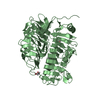
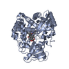
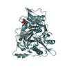
Method to determine structure: MAD / Resolution: 1.8→30.056 Å / Cor.coef. Fo:Fc: 0.972 / Cor.coef. Fo:Fc free: 0.961 / Occupancy max: 1 / Occupancy min: 0.37 / SU B: 2.871 / SU ML: 0.048 / TLS residual ADP flag: LIKELY RESIDUAL / Cross valid method: THROUGHOUT / σ(F): 0 / ESU R: 0.08 / ESU R Free: 0.08 Stereochemistry target values: MAXIMUM LIKELIHOOD WITH PHASES Details: (1). HYDROGENS HAVE BEEN ADDED IN THE RIDING POSITIONS. (2). A MET-INHIBITION PROTOCOL WAS USED FOR SELENOMETHIONINE INCORPORATION DURING PROTEIN EXPRESSION. THE OCCUPANCY OF THE SE ATOMS IN ...Details: (1). HYDROGENS HAVE BEEN ADDED IN THE RIDING POSITIONS. (2). A MET-INHIBITION PROTOCOL WAS USED FOR SELENOMETHIONINE INCORPORATION DURING PROTEIN EXPRESSION. THE OCCUPANCY OF THE SE ATOMS IN THE MSE RESIDUES WAS REDUCED TO 0.75 TO ACCOUNT FOR THE REDUCED SCATTERING POWER DUE TO PARTIAL S-MET INCORPORATION. (3). ATOM RECORD CONTAINS RESIDUAL B FACTORS ONLY. (4). THE DENSITIES OF RESIDUES 27-28, 59-63 AND 286-297 WERE POOR AND THEY WERE NOT MODELED. (5). THE DENSITIES OF RESIDUES 55-58 WERE POOR AND THEIR MODELS ARE NOT RELIABLE. (6). ONE FMN MONOMER WAS MODELED BASED ON THE PRESENCE OF CLEAR AND CONCLUSIVE ELECTRON DENSITY. (7). CA ION, CL IONS, ACETATE (ACT) IONS AND THE R-FORM (PGR) OF 1,2-PROPANE DIOL MOLECULES WERE MODELED BASED ON CRYSTALLIZATION CONDITION.
Rfactor
Num. reflection
% reflection
Selection details
Rfree
0.163
3480
5.1 %
RANDOM
Rwork
0.137
-
-
-
obs
0.138
68745
99.72 %
-
Solvent computation
Ion probe radii: 0.8 Å / Shrinkage radii: 0.8 Å / VDW probe radii: 1.2 Å / Solvent model: MASK
In the structure databanks used in Yorodumi, some data are registered as the other names, "COVID-19 virus" and "2019-nCoV". Here are the details of the virus and the list of structure data.
Jan 31, 2019. EMDB accession codes are about to change! (news from PDBe EMDB page)
EMDB accession codes are about to change! (news from PDBe EMDB page)
The allocation of 4 digits for EMDB accession codes will soon come to an end. Whilst these codes will remain in use, new EMDB accession codes will include an additional digit and will expand incrementally as the available range of codes is exhausted. The current 4-digit format prefixed with “EMD-” (i.e. EMD-XXXX) will advance to a 5-digit format (i.e. EMD-XXXXX), and so on. It is currently estimated that the 4-digit codes will be depleted around Spring 2019, at which point the 5-digit format will come into force.
The EM Navigator/Yorodumi systems omit the EMD- prefix.
Related info.:Q: What is EMD? / ID/Accession-code notation in Yorodumi/EM Navigator
Yorodumi is a browser for structure data from EMDB, PDB, SASBDB, etc.
This page is also the successor to EM Navigator detail page, and also detail information page/front-end page for Omokage search.
The word "yorodu" (or yorozu) is an old Japanese word meaning "ten thousand". "mi" (miru) is to see.
Related info.:EMDB / PDB / SASBDB / Comparison of 3 databanks / Yorodumi Search / Aug 31, 2016. New EM Navigator & Yorodumi / Yorodumi Papers / Jmol/JSmol / Function and homology information / Changes in new EM Navigator and Yorodumi
 Movie
Movie Controller

 Yorodumi
 Open data
 Basic information
 Components
 Keywords
 Function and homology information
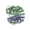
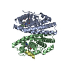
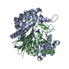
 Anabaena variabilis ATCC 29413 (bacteria)
 X-RAY DIFFRACTION /
  Authors
 Citation
 Structure visualization
 Downloads & links
 Other downloads
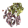


 PDBj
PDBj Assembly
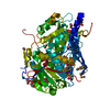


 Mass: 456.344 Da / Num. of mol.: 1 / Source method: obtained synthetically / Formula: C17H21N4O9P
 Mass: 40.078 Da / Num. of mol.: 1 / Source method: obtained synthetically / Formula: Ca
 Mass: 35.453 Da / Num. of mol.: 2 / Source method: obtained synthetically / Formula: Cl
 Mass: 59.044 Da / Num. of mol.: 12 / Source method: obtained synthetically / Formula: C2H3O2
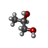
 Mass: 76.094 Da / Num. of mol.: 2 / Source method: obtained synthetically / Formula: C3H8O2
 Sample preparation
 / Beamline: BL9-1 / Wavelength: 0.918370,0.978985,0.979346
 Processing