 Movie
Movie Controller

[English] 日本語
 Yorodumi
- PDB-6bav: Crystal Structure of GltPh R397C in complex with S-Benzyl-L-Cysteine -
+ Open data

- Basic information
| Entry | Database: PDB / ID: 6bav | |||||||||
|---|---|---|---|---|---|---|---|---|---|---|
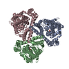
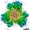
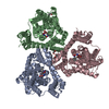
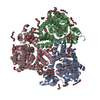
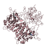
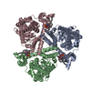
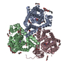
| Title | Crystal Structure of GltPh R397C in complex with S-Benzyl-L-Cysteine | |||||||||
 Components Components | Glutamate transporter homolog | |||||||||
 Keywords Keywords | TRANSPORT PROTEIN / Amino Acid transporter / Transmembrane transporter Aspartate transporter | |||||||||
| Function / homology |  Function and homology information Function and homology informationL-aspartate transmembrane transport / L-aspartate transmembrane transporter activity / L-aspartate import across plasma membrane / amino acid:sodium symporter activity / chloride transmembrane transporter activity / protein homotrimerization / chloride transmembrane transport / metal ion binding / identical protein binding / plasma membrane Similarity search - Function | |||||||||
| Biological species |   Pyrococcus horikoshii (archaea) Pyrococcus horikoshii (archaea) | |||||||||
| Method |  X-RAY DIFFRACTION / SYNCHROTRON / MOLECULAR REPLACEMENT / Resolution: 3.7 Å X-RAY DIFFRACTION / SYNCHROTRON / MOLECULAR REPLACEMENT / Resolution: 3.7 Å | |||||||||
 Authors Authors | Font, J. / Scopelliti, A.J. / Vandenberg, R.J. / Boudker, O. / Ryan, R.M. | |||||||||
| Funding support |  Australia, Australia,  United States, 2items United States, 2items
| |||||||||
 Citation Citation | Journal: Nat Commun / Year: 2018 Title: Structural characterisation reveals insights into substrate recognition by the glutamine transporter ASCT2/SLC1A5. Authors: Scopelliti, A.J. / Font, J. / Vandenberg, R.J. / Boudker, O. / Ryan, R.M. | |||||||||
| History |
|
- Structure visualization
| Structure viewer | Molecule: MolmilJmol/JSmol |
|---|
- Downloads & links
-Download
| PDBx/mmCIF format | 6bav.cif.gz | 466.2 KB | Display | PDBx/mmCIF format |
|---|---|---|---|---|
| PDB format | pdb6bav.ent.gz | 391.7 KB | Display | PDB format |
| PDBx/mmJSON format | 6bav.json.gz | Tree view | PDBx/mmJSON format | |
| Others |  Other downloads Other downloads |
-Validation report
| Arichive directory | https://data.pdbj.org/pub/pdb/validation_reports/ba/6bavftp://data.pdbj.org/pub/pdb/validation_reports/ba/6bav | HTTPS FTP |
|---|
-Related structure data
| Related structure data |  6batC  6bauC  6bmiC  2nwwS S: Starting model for refinement C: citing same article ( |
|---|---|
| Similar structure data |


-Links
 PDBj
PDBj
- Assembly
| Deposited unit | 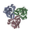
| ||||||||||||||||||||||||||||||||||||
|---|---|---|---|---|---|---|---|---|---|---|---|---|---|---|---|---|---|---|---|---|---|---|---|---|---|---|---|---|---|---|---|---|---|---|---|---|---|
| 1 |
| ||||||||||||||||||||||||||||||||||||
| Unit cell |
| ||||||||||||||||||||||||||||||||||||
| Noncrystallographic symmetry (NCS) | NCS domain:
NCS domain segments:
|
-Components
| #1: Protein | Mass: 44256.660 Da / Num. of mol.: 3 / Mutation: R397C Source method: isolated from a genetically manipulated source Source: (gene. exp.) Pyrococcus horikoshii (archaea)Strain: ATCC 700860 / DSM 12428 / JCM 9974 / NBRC 100139 / OT-3 Gene: PH1295 / Plasmid: PBAD24 / Production host:  #2: Chemical |   Mass: 22.990 Da / Num. of mol.: 3 / Source method: obtained synthetically / Formula: Na Mass: 22.990 Da / Num. of mol.: 3 / Source method: obtained synthetically / Formula: Na#3: Chemical |   Type: L-peptide linking / Mass: 211.281 Da / Num. of mol.: 3 / Source method: obtained synthetically / Formula: C10H13NO2S Type: L-peptide linking / Mass: 211.281 Da / Num. of mol.: 3 / Source method: obtained synthetically / Formula: C10H13NO2S |
|---|
-Experimental details
-Experiment
| Experiment | Method: X-RAY DIFFRACTION / Number of used crystals: 1 |
|---|
- Sample preparation
| Crystal | Density Matthews: 4.74 Å3/Da / Density % sol: 74.03 % / Description: Sword |
|---|---|
| Crystal grow | Temperature: 280 K / Method: vapor diffusion, hanging drop / pH: 7 Details: 50 mM citric acid 50 mM Disodium phosphate 100 mM Lithium sulfate 15-23% PEG1000 |
-Data collection
| Diffraction | Mean temperature: 100 K |
|---|---|
| Diffraction source | Source: SYNCHROTRON / Site: Australian Synchrotron / Beamline: MX2 / Wavelength: 0.9537 Å |
| Detector | Type: ADSC QUANTUM 315r / Detector: CCD / Date: Sep 27, 2014 |
| Radiation | Protocol: SINGLE WAVELENGTH / Monochromatic (M) / Laue (L): M / Scattering type: x-ray |
| Radiation wavelength | Wavelength: 0.9537 Å / Relative weight: 1 |
| Reflection | Resolution: 3.7→49.07 Å / Num. obs: 24809 / % possible obs: 97.5 % / Redundancy: 3.5 % / CC1/2: 0.997 / Rmerge(I) obs: 0.078 / Rpim(I) all: 0.046 / Rrim(I) all: 0.091 / Net I/σ(I): 8.7 |
| Reflection shell | Resolution: 3.7→3.9 Å / Redundancy: 3.4 % / Rmerge(I) obs: 0.552 / Mean I/σ(I) obs: 1.7 / Num. unique obs: 12538 / CC1/2: 0.833 / Rpim(I) all: 0.335 / Rrim(I) all: 0.653 / % possible all: 98.9 |
- Processing
| Software |
| ||||||||||||||||||||||||||||||||||||||||||||||||||||||||||||||||||||||||||||||||||||||||||||||||||||
|---|---|---|---|---|---|---|---|---|---|---|---|---|---|---|---|---|---|---|---|---|---|---|---|---|---|---|---|---|---|---|---|---|---|---|---|---|---|---|---|---|---|---|---|---|---|---|---|---|---|---|---|---|---|---|---|---|---|---|---|---|---|---|---|---|---|---|---|---|---|---|---|---|---|---|---|---|---|---|---|---|---|---|---|---|---|---|---|---|---|---|---|---|---|---|---|---|---|---|---|---|---|
| Refinement | Method to determine structure: MOLECULAR REPLACEMENT Starting model: 2NWW Resolution: 3.7→40 Å / Cor.coef. Fo:Fc: 0.904 / Cor.coef. Fo:Fc free: 0.93 / SU B: 93.68 / SU ML: 0.566 / Cross valid method: THROUGHOUT / σ(F): 0 / ESU R Free: 0.611 / Stereochemistry target values: MAXIMUM LIKELIHOOD Details: HYDROGENS HAVE BEEN ADDED IN THE RIDING POSITIONS U VALUES : WITH TLS ADDED
| ||||||||||||||||||||||||||||||||||||||||||||||||||||||||||||||||||||||||||||||||||||||||||||||||||||
| Solvent computation | Ion probe radii: 0.8 Å / Shrinkage radii: 0.8 Å / VDW probe radii: 1.4 Å / Solvent model: MASK | ||||||||||||||||||||||||||||||||||||||||||||||||||||||||||||||||||||||||||||||||||||||||||||||||||||
| Displacement parameters | Biso max: 325.11 Å2 / Biso mean: 184.509 Å2 / Biso min: 120.59 Å2
| ||||||||||||||||||||||||||||||||||||||||||||||||||||||||||||||||||||||||||||||||||||||||||||||||||||
| Refinement step | Cycle: final / Resolution: 3.7→40 Å
| ||||||||||||||||||||||||||||||||||||||||||||||||||||||||||||||||||||||||||||||||||||||||||||||||||||
| Refine LS restraints |
| ||||||||||||||||||||||||||||||||||||||||||||||||||||||||||||||||||||||||||||||||||||||||||||||||||||
| Refine LS restraints NCS | Ens-ID: 1 / Refine-ID: X-RAY DIFFRACTION
| ||||||||||||||||||||||||||||||||||||||||||||||||||||||||||||||||||||||||||||||||||||||||||||||||||||
| LS refinement shell | Resolution: 3.7→3.795 Å / Rfactor Rfree error: 0 / Total num. of bins used: 20
| ||||||||||||||||||||||||||||||||||||||||||||||||||||||||||||||||||||||||||||||||||||||||||||||||||||
| Refinement TLS params. | Method: refined / Refine-ID: X-RAY DIFFRACTION
| ||||||||||||||||||||||||||||||||||||||||||||||||||||||||||||||||||||||||||||||||||||||||||||||||||||
| Refinement TLS group |
|
