[English] 日本語
 Yorodumi
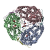
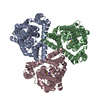
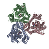
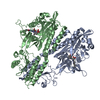
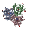
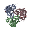
- PDB-6x17: Outward-facing state of the glutamate transporter homologue GltPh... -
+ Open data
- Basic information
| Entry | Database: PDB / ID: 6x17 | |||||||||
|---|---|---|---|---|---|---|---|---|---|---|
| Title | Outward-facing state of the glutamate transporter homologue GltPh in complex with TBOA | |||||||||
 Components Components | Glutamate transporter homologue GltPh | |||||||||
 Keywords Keywords | TRANSPORT PROTEIN / sodium-coupled L-aspartate transporter | |||||||||
| Function / homology |  Function and homology information Function and homology informationL-aspartate transmembrane transport / L-aspartate transmembrane transporter activity / amino acid:sodium symporter activity / L-aspartate import across plasma membrane / chloride transmembrane transporter activity / protein homotrimerization / chloride transmembrane transport / metal ion binding / identical protein binding / plasma membrane Similarity search - Function | |||||||||
| Biological species |   Pyrococcus horikoshii (archaea) Pyrococcus horikoshii (archaea) | |||||||||
| Method | ELECTRON MICROSCOPY / single particle reconstruction / cryo EM / Resolution: 3.66 Å | |||||||||
 Authors Authors | Wang, X. / Boudker, O. | |||||||||
| Funding support |  United States, 2items United States, 2items
| |||||||||
 Citation Citation |  Journal: Elife / Year: 2020 Journal: Elife / Year: 2020Title: Large domain movements through the lipid bilayer mediate substrate release and inhibition of glutamate transporters. Authors: Xiaoyu Wang / Olga Boudker /  Abstract: Glutamate transporters are essential players in glutamatergic neurotransmission in the brain, where they maintain extracellular glutamate below cytotoxic levels and allow for rounds of transmission. ...Glutamate transporters are essential players in glutamatergic neurotransmission in the brain, where they maintain extracellular glutamate below cytotoxic levels and allow for rounds of transmission. The structural bases of their function are well established, particularly within a model archaeal homolog, sodium, and aspartate symporter Glt. However, the mechanism of gating on the cytoplasmic side of the membrane remains ambiguous. We report Cryo-EM structures of Glt reconstituted into nanodiscs, including those structurally constrained in the cytoplasm-facing state and either apo, bound to sodium ions only, substrate, or blockers. The structures show that both substrate translocation and release involve movements of the bulky transport domain through the lipid bilayer. They further reveal a novel mode of inhibitor binding and show how solutes release is coupled to protein conformational changes. Finally, we describe how domain movements are associated with the displacement of bound lipids and significant membrane deformations, highlighting the potential regulatory role of the bilayer. | |||||||||
| History |
|
- Structure visualization
| Movie |
 Movie viewer Movie viewer |
|---|---|
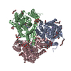
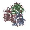
| Structure viewer | Molecule:  Molmil Molmil Jmol/JSmol Jmol/JSmol |
- Downloads & links
- Download
| PDBx/mmCIF format |  6x17.cif.gz 6x17.cif.gz | 207.7 KB | Display |  PDBx/mmCIF format PDBx/mmCIF format |
|---|---|---|---|---|
| PDB format |  pdb6x17.ent.gz pdb6x17.ent.gz | 169.8 KB | Display |  PDB format PDB format |
| PDBx/mmJSON format |  6x17.json.gz 6x17.json.gz | Tree view |  PDBx/mmJSON format PDBx/mmJSON format | |
| Others |  Other downloads Other downloads |
-Validation report
| Arichive directory |  https://data.pdbj.org/pub/pdb/validation_reports/x1/6x17 https://data.pdbj.org/pub/pdb/validation_reports/x1/6x17 ftp://data.pdbj.org/pub/pdb/validation_reports/x1/6x17 ftp://data.pdbj.org/pub/pdb/validation_reports/x1/6x17 | HTTPS FTP |
|---|
-Related structure data
| Related structure data |  21991MC  6x12C  6x13C  6x14C  6x15C  6x16C M: map data used to model this data C: citing same article ( |
|---|---|
| Similar structure data |
- Links
Links
- Assembly
| Deposited unit | 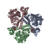
|
|---|---|
| 1 |
|
- Components
| #1: Protein | Mass: 44669.957 Da / Num. of mol.: 3 Source method: isolated from a genetically manipulated source Source: (gene. exp.)   Pyrococcus horikoshii (archaea) / Production host: Pyrococcus horikoshii (archaea) / Production host:  #2: Chemical | #3: Water | ChemComp-HOH / | Has ligand of interest | N | Has protein modification | N | |
|---|
-Experimental details
-Experiment
| Experiment | Method: ELECTRON MICROSCOPY |
|---|---|
| EM experiment | Aggregation state: PARTICLE / 3D reconstruction method: single particle reconstruction |
- Sample preparation
| Component | Name: Complex of GltPh with TBOA in MSP1E3 nanodisc / Type: COMPLEX / Entity ID: #1 / Source: RECOMBINANT |
|---|---|
| Molecular weight | Value: 0.134 MDa / Experimental value: NO |
| Source (natural) | Organism:   Pyrococcus horikoshii (archaea) Pyrococcus horikoshii (archaea) |
| Source (recombinant) | Organism:  |
| Buffer solution | pH: 7.4 |
| Specimen | Embedding applied: YES / Shadowing applied: NO / Staining applied: NO / Vitrification applied: YES |
| Specimen support | Details: unspecified |
| EM embedding | Material: ice |
| Vitrification | Cryogen name: ETHANE |
- Electron microscopy imaging
| Experimental equipment |  Model: Titan Krios / Image courtesy: FEI Company |
|---|---|
| Microscopy | Model: FEI TITAN KRIOS |
| Electron gun | Electron source:  FIELD EMISSION GUN / Accelerating voltage: 300 kV / Illumination mode: FLOOD BEAM FIELD EMISSION GUN / Accelerating voltage: 300 kV / Illumination mode: FLOOD BEAM |
| Electron lens | Mode: BRIGHT FIELD |
| Image recording | Electron dose: 69.7 e/Å2 / Film or detector model: GATAN K3 (6k x 4k) |
- Processing
| CTF correction | Type: PHASE FLIPPING AND AMPLITUDE CORRECTION |
|---|---|
| Symmetry | Point symmetry: C3 (3 fold cyclic) |
| 3D reconstruction | Resolution: 3.66 Å / Resolution method: FSC 0.143 CUT-OFF / Num. of particles: 88961 / Symmetry type: POINT |
 Movie
Movie Controller



 PDBj
PDBj
