 Movie
Movie Controller

[English] 日本語
 Yorodumi
- PDB-3gcm: Crystal Structure of E. coli polynucleotide phosphorylase bound t... -
+ Open data

- Basic information
| Entry | Database: PDB / ID: 3gcm | ||||||
|---|---|---|---|---|---|---|---|
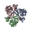
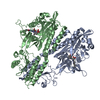
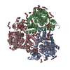
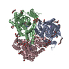
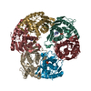
| Title | Crystal Structure of E. coli polynucleotide phosphorylase bound to RNA and RNase E | ||||||
 Components Components |
| ||||||
 Keywords Keywords | transferase / protein binding / Protein-RNA complex / Cytoplasm / Nucleotidyltransferase / RNA-binding / Transferase / Hydrolase / transferase - protein binding COMPLEX | ||||||
| Function / homology |  Function and homology information Function and homology informationribonuclease E / ribonuclease E activity / polyribonucleotide nucleotidyltransferase / polyribonucleotide nucleotidyltransferase activity / tRNA processing / mRNA catabolic process / RNA processing / cytoplasmic side of plasma membrane / rRNA processing / 3'-5'-RNA exonuclease activity ...ribonuclease E / ribonuclease E activity / polyribonucleotide nucleotidyltransferase / polyribonucleotide nucleotidyltransferase activity / tRNA processing / mRNA catabolic process / RNA processing / cytoplasmic side of plasma membrane / rRNA processing / 3'-5'-RNA exonuclease activity / tRNA binding / rRNA binding / magnesium ion binding / RNA binding / zinc ion binding / cytoplasm / cytosol Similarity search - Function | ||||||
| Biological species |  | ||||||
| Method |  X-RAY DIFFRACTION / SYNCHROTRON / MOLECULAR REPLACEMENT / Resolution: 2.5 Å X-RAY DIFFRACTION / SYNCHROTRON / MOLECULAR REPLACEMENT / Resolution: 2.5 Å | ||||||
 Authors Authors | Nurmohamed, S. / Luisi, B.L. | ||||||
 Citation Citation | Journal: J.Mol.Biol. / Year: 2009 Title: Crystal structure of Escherichia coli polynucleotide phosphorylase core bound to RNase E, RNA and manganese: implications for catalytic mechanism and RNA degradosome assembly. Authors: Nurmohamed, S. / Vaidialingam, B. / Callaghan, A.J. / Luisi, B.F. | ||||||
| History |
|
- Structure visualization
| Structure viewer | Molecule: MolmilJmol/JSmol |
|---|
- Downloads & links
-Download
| PDBx/mmCIF format | 3gcm.cif.gz | 350.1 KB | Display | PDBx/mmCIF format |
|---|---|---|---|---|
| PDB format | pdb3gcm.ent.gz | 281.1 KB | Display | PDB format |
| PDBx/mmJSON format | 3gcm.json.gz | Tree view | PDBx/mmJSON format | |
| Others |  Other downloads Other downloads |
-Validation report
| Arichive directory | https://data.pdbj.org/pub/pdb/validation_reports/gc/3gcmftp://data.pdbj.org/pub/pdb/validation_reports/gc/3gcm | HTTPS FTP |
|---|
-Related structure data
| Related structure data |  3gllC  3gmeC 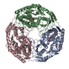 3h1cC  1e3hS C: citing same article ( S: Starting model for refinement |
|---|---|
| Similar structure data |


-Links
 PDBj
PDBj


- Assembly
| Deposited unit | 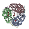
| ||||||||
|---|---|---|---|---|---|---|---|---|---|
| 1 |
| ||||||||
| Unit cell |
| ||||||||
| Components on special symmetry positions |
|
-Components
-Protein / Protein/peptide , 2 types, 6 molecules ABCDEF
| #1: Protein | Mass: 59657.812 Da / Num. of mol.: 3 / Fragment: residues 1-549 / Source method: isolated from a natural source / Source: (natural) References: UniProt: A7ZS61, polyribonucleotide nucleotidyltransferase #2: Protein/peptide | Mass: 4340.687 Da / Num. of mol.: 3 Fragment: RNase E recognition microdomain, residues 1021-1061 Source method: obtained synthetically / Details: This sequence occurs naturally in E. coli References: UniProt: A7ZKI9, Hydrolases; Acting on ester bonds; Phosphoric-diester hydrolases |
|---|
-Non-polymers , 4 types, 898 molecules 


| #3: Chemical | ChemComp-FLC /  Mass: 189.100 Da / Num. of mol.: 8 / Source method: obtained synthetically / Formula: C6H5O7 Mass: 189.100 Da / Num. of mol.: 8 / Source method: obtained synthetically / Formula: C6H5O7#4: Chemical |  Mass: 24.305 Da / Num. of mol.: 3 / Source method: obtained synthetically / Formula: Mg Mass: 24.305 Da / Num. of mol.: 3 / Source method: obtained synthetically / Formula: Mg#5: Chemical |  Mass: 363.221 Da / Num. of mol.: 3 / Source method: obtained synthetically / Formula: C10H14N5O8P Mass: 363.221 Da / Num. of mol.: 3 / Source method: obtained synthetically / Formula: C10H14N5O8P#6: Water | ChemComp-HOH / | Mass: 18.015 Da / Num. of mol.: 884 / Source method: isolated from a natural source / Formula: H2O |
|---|
-Experimental details
-Experiment
| Experiment | Method: X-RAY DIFFRACTION / Number of used crystals: 1 |
|---|
- Sample preparation
| Crystal | Density Matthews: 3.84 Å3/Da / Density % sol: 67.96 % |
|---|---|
| Crystal grow | Temperature: 293.15 K / Method: vapor diffusion, hanging drop / pH: 4.5 Details: 0.2 M ammonium hydrogen citrate, 17 % w/v PEG 3350, pH 4.5, VAPOR DIFFUSION, HANGING DROP, temperature 293.15K |
-Data collection
| Diffraction | Mean temperature: 120 K |
|---|---|
| Diffraction source | Source: SYNCHROTRON / Site: ESRF  / Beamline: ID23-2 / Wavelength: 0.873 Å / Beamline: ID23-2 / Wavelength: 0.873 Å |
| Detector | Type: MARMOSAIC 225 mm CCD / Detector: CCD / Date: Oct 4, 2006 / Details: Monochromator |
| Radiation | Monochromator: horizontally diffracting silicon / Protocol: SINGLE WAVELENGTH / Monochromatic (M) / Laue (L): M / Scattering type: x-ray |
| Radiation wavelength | Wavelength: 0.873 Å / Relative weight: 1 |
| Reflection | Resolution: 2.5→20 Å / Num. all: 102846 / Num. obs: 97645 / % possible obs: 99.3 % / Observed criterion σ(F): 2 / Observed criterion σ(I): 2 / Redundancy: 6.6 % / Biso Wilson estimate: 27.176 Å2 |
| Reflection shell | Resolution: 2.5→2.59 Å / Rmerge(I) obs: 0.619 / Mean I/σ(I) obs: 2.2 / % possible all: 99.5 |
- Processing
| Software |
| ||||||||||||||||||||||||||||||||||||||||||||||||||||||||||||||||||||||||||||||||||||||||||||||||||||||||||||||||||||||||||||||||||||||||||||||||||||||||||||||||||||||||||
|---|---|---|---|---|---|---|---|---|---|---|---|---|---|---|---|---|---|---|---|---|---|---|---|---|---|---|---|---|---|---|---|---|---|---|---|---|---|---|---|---|---|---|---|---|---|---|---|---|---|---|---|---|---|---|---|---|---|---|---|---|---|---|---|---|---|---|---|---|---|---|---|---|---|---|---|---|---|---|---|---|---|---|---|---|---|---|---|---|---|---|---|---|---|---|---|---|---|---|---|---|---|---|---|---|---|---|---|---|---|---|---|---|---|---|---|---|---|---|---|---|---|---|---|---|---|---|---|---|---|---|---|---|---|---|---|---|---|---|---|---|---|---|---|---|---|---|---|---|---|---|---|---|---|---|---|---|---|---|---|---|---|---|---|---|---|---|---|---|---|---|---|
| Refinement | Method to determine structure: MOLECULAR REPLACEMENT Starting model: 1E3H Resolution: 2.5→20 Å / Cor.coef. Fo:Fc: 0.951 / Cor.coef. Fo:Fc free: 0.917 / SU B: 5.883 / SU ML: 0.133 / Cross valid method: THROUGHOUT / ESU R: 0.244 / ESU R Free: 0.212 / Stereochemistry target values: MAXIMUM LIKELIHOOD / Details: HYDROGENS HAVE BEEN ADDED IN THE RIDING POSITIONS
| ||||||||||||||||||||||||||||||||||||||||||||||||||||||||||||||||||||||||||||||||||||||||||||||||||||||||||||||||||||||||||||||||||||||||||||||||||||||||||||||||||||||||||
| Solvent computation | Ion probe radii: 0.8 Å / Shrinkage radii: 0.8 Å / VDW probe radii: 1.2 Å / Solvent model: MASK | ||||||||||||||||||||||||||||||||||||||||||||||||||||||||||||||||||||||||||||||||||||||||||||||||||||||||||||||||||||||||||||||||||||||||||||||||||||||||||||||||||||||||||
| Displacement parameters | Biso mean: 27.176 Å2
| ||||||||||||||||||||||||||||||||||||||||||||||||||||||||||||||||||||||||||||||||||||||||||||||||||||||||||||||||||||||||||||||||||||||||||||||||||||||||||||||||||||||||||
| Refinement step | Cycle: LAST / Resolution: 2.5→20 Å
| ||||||||||||||||||||||||||||||||||||||||||||||||||||||||||||||||||||||||||||||||||||||||||||||||||||||||||||||||||||||||||||||||||||||||||||||||||||||||||||||||||||||||||
| Refine LS restraints |
| ||||||||||||||||||||||||||||||||||||||||||||||||||||||||||||||||||||||||||||||||||||||||||||||||||||||||||||||||||||||||||||||||||||||||||||||||||||||||||||||||||||||||||
| LS refinement shell | Resolution: 2.499→2.563 Å / Total num. of bins used: 20
|
