 Movie
Movie Controller

[English] 日本語
 Yorodumi
- PDB-3h1c: Crystal structure of Polynucleotide Phosphorylase (PNPase) core b... -
+ Open data

- Basic information
| Entry | Database: PDB / ID: 3h1c | ||||||
|---|---|---|---|---|---|---|---|
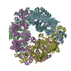
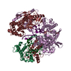
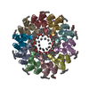
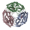
| Title | Crystal structure of Polynucleotide Phosphorylase (PNPase) core bound to RNase E and Tungstate | ||||||
 Components Components |
| ||||||
 Keywords Keywords | TRANSFERASE / polynucleotide phosphorylase / RNA turnover / Cytoplasm / Nucleotidyltransferase / RNA-binding / Stress response / Endonuclease / Hydrolase / Nuclease | ||||||
| Function / homology |  Function and homology information Function and homology informationrRNA 5'-end processing / bacterial degradosome / ribonuclease E / ribonuclease E activity / polyribonucleotide nucleotidyltransferase / polyribonucleotide nucleotidyltransferase activity / endoribonuclease complex / DEAD/H-box RNA helicase binding / 7S RNA binding / cyclic-di-GMP binding ...rRNA 5'-end processing / bacterial degradosome / ribonuclease E / ribonuclease E activity / polyribonucleotide nucleotidyltransferase / polyribonucleotide nucleotidyltransferase activity / endoribonuclease complex / DEAD/H-box RNA helicase binding / 7S RNA binding / cyclic-di-GMP binding / RNA catabolic process / tRNA processing / mRNA catabolic process / protein complex oligomerization / RNA nuclease activity / RNA processing / RNA endonuclease activity / cytoplasmic side of plasma membrane / rRNA processing / response to heat / 3'-5'-RNA exonuclease activity / molecular adaptor activity / protein homotetramerization / tRNA binding / rRNA binding / magnesium ion binding / RNA binding / zinc ion binding / membrane / identical protein binding / cytoplasm / cytosol Similarity search - Function | ||||||
| Biological species |  | ||||||
| Method |  X-RAY DIFFRACTION / SYNCHROTRON / MOLECULAR REPLACEMENT / Resolution: 3.57 Å X-RAY DIFFRACTION / SYNCHROTRON / MOLECULAR REPLACEMENT / Resolution: 3.57 Å | ||||||
 Authors Authors | Nurmohamed, S. | ||||||
 Citation Citation | Journal: J.Mol.Biol. / Year: 2009 Title: Crystal structure of Escherichia coli polynucleotide phosphorylase core bound to RNase E, RNA and manganese: implications for catalytic mechanism and RNA degradosome assembly Authors: Nurmohamed, S. / Vaidialingam, B. / Callaghan, A.J. / Luisi, B.F. | ||||||
| History |
|
- Structure visualization
| Structure viewer | Molecule: MolmilJmol/JSmol |
|---|
- Downloads & links
-Download
| PDBx/mmCIF format | 3h1c.cif.gz | 1.2 MB | Display | PDBx/mmCIF format |
|---|---|---|---|---|
| PDB format | pdb3h1c.ent.gz | 1021.8 KB | Display | PDB format |
| PDBx/mmJSON format | 3h1c.json.gz | Tree view | PDBx/mmJSON format | |
| Others |  Other downloads Other downloads |
-Validation report
| Arichive directory | https://data.pdbj.org/pub/pdb/validation_reports/h1/3h1cftp://data.pdbj.org/pub/pdb/validation_reports/h1/3h1c | HTTPS FTP |
|---|
-Related structure data
| Related structure data | 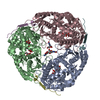 3gcmSC  3gllC  3gmeC S: Starting model for refinement C: citing same article ( |
|---|---|
| Similar structure data |


-Links
 PDBj
PDBj


- Assembly


-Components
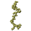
| #1: Protein | Mass: 59656.828 Da / Num. of mol.: 12 / Source method: isolated from a natural source / Source: (natural) References: UniProt: P05055, polyribonucleotide nucleotidyltransferase #2: Protein/peptide | Mass: 4340.687 Da / Num. of mol.: 12 / Fragment: UNP residues 1021-1061 / Source method: obtained synthetically / Details: This sequence occurs naturally in E. coli / References: UniProt: P21513, ribonuclease E #3: Chemical | ChemComp-WO4 /   Mass: 247.838 Da / Num. of mol.: 21 / Source method: obtained synthetically / Formula: WO4 Mass: 247.838 Da / Num. of mol.: 21 / Source method: obtained synthetically / Formula: WO4 |
|---|
-Experimental details
-Experiment
| Experiment | Method: X-RAY DIFFRACTION / Number of used crystals: 1 |
|---|
- Sample preparation
| Crystal | Density Matthews: 3.79 Å3/Da / Density % sol: 67.56 % |
|---|---|
| Crystal grow | Temperature: 291 K / Method: vapor diffusion, hanging drop / pH: 4.5 Details: 0.2M ammonium hydrogen citrate, 17% PEG 3350, 50mM disodium tungstate, pH4.5, VAPOR DIFFUSION, HANGING DROP, temperature 291K |
-Data collection
| Diffraction | Mean temperature: 100 K |
|---|---|
| Diffraction source | Source: SYNCHROTRON / Site: ESRF  / Beamline: ID23-2 / Wavelength: 0.873 Å / Beamline: ID23-2 / Wavelength: 0.873 Å |
| Detector | Type: MARMOSAIC 225 mm CCD / Detector: CCD / Date: Jul 15, 2006 / Details: Monochromator |
| Radiation | Protocol: SINGLE WAVELENGTH / Monochromatic (M) / Laue (L): M / Scattering type: x-ray |
| Radiation wavelength | Wavelength: 0.873 Å / Relative weight: 1 |
| Reflection | Resolution: 3.57→79.5 Å / Num. all: 129998 / Num. obs: 123425 / % possible obs: 94.2 % / Observed criterion σ(F): 2 / Observed criterion σ(I): 2 / Redundancy: 2.8 % / Biso Wilson estimate: 57.1 Å2 / Rmerge(I) obs: 0.123 / Net I/σ(I): 11.4 |
| Reflection shell | Resolution: 3.57→3.73 Å / Redundancy: 2.4 % / Rmerge(I) obs: 0.392 / Mean I/σ(I) obs: 2.3 / Num. unique all: 129998 / % possible all: 94.2 |
- Processing
| Software |
| |||||||||||||||||||||||||
|---|---|---|---|---|---|---|---|---|---|---|---|---|---|---|---|---|---|---|---|---|---|---|---|---|---|---|
| Refinement | Method to determine structure: MOLECULAR REPLACEMENT Starting model: PDB ENTRY 3GCM Resolution: 3.57→25 Å / Cross valid method: THROUGHOUT / σ(F): 2 / σ(I): 2
| |||||||||||||||||||||||||
| Refinement step | Cycle: LAST / Resolution: 3.57→25 Å
|
