| Entry | Database: PDB / ID: 5xex
|
|---|
| Title | Crystal structure of S.aureus PNPase catalytic domain |
|---|
 Components Components | Polyribonucleotide nucleotidyltransferase |
|---|
 Keywords Keywords | TRANSFERASE / polynucleotide phosphorylase / catalytic domain |
|---|
| Function / homology |  Function and homology information Function and homology information
polyribonucleotide nucleotidyltransferase / polyribonucleotide nucleotidyltransferase activity / RNA catabolic process / mRNA catabolic process / RNA processing / 3'-5'-RNA exonuclease activity / magnesium ion binding / RNA binding / cytosolSimilarity search - Function Polyribonucleotide nucleotidyltransferase, RNA-binding domain superfamily / Polyribonucleotide nucleotidyltransferase / Polyribonucleotide nucleotidyltransferase, RNA-binding domain / Polyribonucleotide nucleotidyltransferase, RNA binding domain / GHMP Kinase, N-terminal domain / Exoribonuclease, phosphorolytic domain 2 / 3' exoribonuclease family, domain 2 / Exoribonuclease, phosphorolytic domain 1 / PNPase/RNase PH domain superfamily / Exoribonuclease, PH domain 2 superfamily ...Polyribonucleotide nucleotidyltransferase, RNA-binding domain superfamily / Polyribonucleotide nucleotidyltransferase / Polyribonucleotide nucleotidyltransferase, RNA-binding domain / Polyribonucleotide nucleotidyltransferase, RNA binding domain / GHMP Kinase, N-terminal domain / Exoribonuclease, phosphorolytic domain 2 / 3' exoribonuclease family, domain 2 / Exoribonuclease, phosphorolytic domain 1 / PNPase/RNase PH domain superfamily / Exoribonuclease, PH domain 2 superfamily / 3' exoribonuclease family, domain 1 / KH domain / K Homology domain, type 1 / Type-1 KH domain profile. / K Homology domain, type 1 superfamily / Ribosomal Protein S5; domain 2 / S1 domain profile. / Ribosomal protein S1-like RNA-binding domain / S1 RNA binding domain / S1 domain / K Homology domain / K homology RNA-binding domain / Ribosomal protein S5 domain 2-type fold / Nucleic acid-binding, OB-fold / 2-Layer Sandwich / Alpha BetaSimilarity search - Domain/homology |
|---|
| Biological species |   Staphylococcus aureus (bacteria) Staphylococcus aureus (bacteria) |
|---|
| Method |  X-RAY DIFFRACTION / SYNCHROTRON / MOLECULAR REPLACEMENT / Resolution: 2.2 Å X-RAY DIFFRACTION / SYNCHROTRON / MOLECULAR REPLACEMENT / Resolution: 2.2 Å |
|---|
 Authors Authors | Wang, X. / Zhang, X. / Zang, J. |
|---|
 Citation Citation | Journal: FEBS Lett. / Year: 2017
Title: Enolase binds to RnpA in competition with PNPase in Staphylococcus aureus
Authors: Wang, X. / Wang, C. / Wu, M. / Tian, T. / Cheng, T. / Zhang, X. / Zang, J. |
|---|
| History | | Deposition | Apr 6, 2017 | Deposition site: PDBJ / Processing site: PDBJ |
|---|
| Revision 1.0 | Oct 18, 2017 | Provider: repository / Type: Initial release |
|---|
| Revision 1.1 | Nov 29, 2017 | Group: Database references / Category: citation
Item: _citation.journal_volume / _citation.page_first / _citation.page_last |
|---|
| Revision 1.2 | Feb 7, 2018 | Group: Database references / Category: pdbx_related_exp_data_set |
|---|
| Revision 1.3 | Nov 22, 2023 | Group: Data collection / Database references / Refinement description
Category: chem_comp_atom / chem_comp_bond ...chem_comp_atom / chem_comp_bond / database_2 / pdbx_initial_refinement_model
Item: _database_2.pdbx_DOI / _database_2.pdbx_database_accession |
|---|
|
|---|
 Movie
Movie Controller


 Open data
 Basic information
 Structure visualization
 Downloads & links
 Other downloads
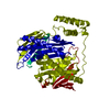


 PDBj
PDBj


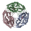
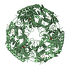
 Assembly


 Mass: 106.120 Da / Num. of mol.: 6 / Source method: obtained synthetically / Formula: C4H10O3
 Mass: 92.094 Da / Num. of mol.: 6 / Source method: obtained synthetically / Formula: C3H8O3
 Mass: 177.975 Da / Num. of mol.: 6 / Source method: obtained synthetically / Formula: H4O7P2
 Mass: 18.015 Da / Num. of mol.: 242 / Source method: isolated from a natural source / Formula: H2O
 Sample preparation
 / Beamline: BL19U1 / Wavelength: 0.97846 Å
 Processing