 Movie
Movie Controller

[English] 日本語
 Yorodumi
- PDB-4aid: Crystal structure of C. crescentus PNPase bound to RNase E recogn... -
+ Open data

- Basic information
| Entry | Database: PDB / ID: 4aid | ||||||
|---|---|---|---|---|---|---|---|
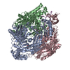
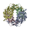
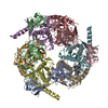
| Title | Crystal structure of C. crescentus PNPase bound to RNase E recognition peptide | ||||||
 Components Components |
| ||||||
 Keywords Keywords | TRANSFERASE/PEPTIDE / TRANSFERASE-PEPTIDE COMPLEX | ||||||
| Function / homology |  Function and homology information Function and homology informationribonuclease E / ribonuclease E activity / polyribonucleotide nucleotidyltransferase / polyribonucleotide nucleotidyltransferase activity / tRNA processing / mRNA catabolic process / RNA processing / cytoplasmic side of plasma membrane / rRNA processing / 3'-5'-RNA exonuclease activity ...ribonuclease E / ribonuclease E activity / polyribonucleotide nucleotidyltransferase / polyribonucleotide nucleotidyltransferase activity / tRNA processing / mRNA catabolic process / RNA processing / cytoplasmic side of plasma membrane / rRNA processing / 3'-5'-RNA exonuclease activity / tRNA binding / rRNA binding / magnesium ion binding / RNA binding / zinc ion binding / cytoplasm / cytosol Similarity search - Function | ||||||
| Biological species |  CAULOBACTER VIBRIOIDES (bacteria) CAULOBACTER VIBRIOIDES (bacteria) | ||||||
| Method |  X-RAY DIFFRACTION / SYNCHROTRON / MOLECULAR REPLACEMENT / Resolution: 2.6 Å X-RAY DIFFRACTION / SYNCHROTRON / MOLECULAR REPLACEMENT / Resolution: 2.6 Å | ||||||
 Authors Authors | Hardwick, S.W. / Gubbey, T. / Hug, I. / Jenal, U. / Luisi, B.F. | ||||||
 Citation Citation | Journal: Open Biol. / Year: 2012 Title: Crystal Structure of Caulobacter Crescentus Polynucleotide Phosphorylase Reveals a Mechanism of RNA Substrate Channelling and RNA Degradosome Assembly. Authors: Hardwick, S.W. / Gubbey, T. / Hug, I. / Jenal, U. / Luisi, B.F. | ||||||
| History |
|
- Structure visualization
| Structure viewer | Molecule: MolmilJmol/JSmol |
|---|
- Downloads & links
-Download
| PDBx/mmCIF format | 4aid.cif.gz | 626.9 KB | Display | PDBx/mmCIF format |
|---|---|---|---|---|
| PDB format | pdb4aid.ent.gz | 516.6 KB | Display | PDB format |
| PDBx/mmJSON format | 4aid.json.gz | Tree view | PDBx/mmJSON format | |
| Others |  Other downloads Other downloads |
-Validation report
| Arichive directory | https://data.pdbj.org/pub/pdb/validation_reports/ai/4aidftp://data.pdbj.org/pub/pdb/validation_reports/ai/4aid | HTTPS FTP |
|---|
-Related structure data
| Related structure data |  4aimC  4am3C  3gmeS S: Starting model for refinement C: citing same article ( |
|---|---|
| Similar structure data |
-Links
 PDBj
PDBj


- Assembly
| Deposited unit | 
| ||||||||
|---|---|---|---|---|---|---|---|---|---|
| 1 | 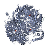
| ||||||||
| 2 | 
| ||||||||
| 3 | 
| ||||||||
| Unit cell |
|
-Components
| #1: Protein | Mass: 78159.367 Da / Num. of mol.: 3 Source method: isolated from a genetically manipulated source Source: (gene. exp.) CAULOBACTER VIBRIOIDES (bacteria) / Strain: CB15 / Production host: References: UniProt: Q9AC32, polyribonucleotide nucleotidyltransferase #2: Protein/peptide | Mass: 1798.081 Da / Num. of mol.: 3 / Fragment: PNPASE BINDING PEPTIDE - GWW, RESIDUES 885-898 / Source method: obtained synthetically / Source: (synth.) CAULOBACTER VIBRIOIDES (bacteria) / References: UniProt: Q9A749#3: Chemical |   Mass: 94.971 Da / Num. of mol.: 3 / Source method: obtained synthetically / Formula: PO4 Mass: 94.971 Da / Num. of mol.: 3 / Source method: obtained synthetically / Formula: PO4#4: Water | ChemComp-HOH / |  Mass: 18.015 Da / Num. of mol.: 201 / Source method: isolated from a natural source / Formula: H2O Mass: 18.015 Da / Num. of mol.: 201 / Source method: isolated from a natural source / Formula: H2O |
|---|
-Experimental details
-Experiment
| Experiment | Method: X-RAY DIFFRACTION / Number of used crystals: 1 |
|---|
- Sample preparation
| Crystal | Density Matthews: 3.01 Å3/Da / Density % sol: 59 % / Description: NONE |
|---|---|
| Crystal grow | Details: 19% WT/V PEG 3350, 0.15 M DL-MALIC ACID |
-Data collection
| Diffraction | Mean temperature: 100 K |
|---|---|
| Diffraction source | Source: SYNCHROTRON / Site: Diamond  / Beamline: I02 / Wavelength: 0.9795 / Beamline: I02 / Wavelength: 0.9795 |
| Detector | Type: ADSC CCD / Detector: CCD / Date: Feb 3, 2011 |
| Radiation | Protocol: SINGLE WAVELENGTH / Monochromatic (M) / Laue (L): M / Scattering type: x-ray |
| Radiation wavelength | Wavelength: 0.9795 Å / Relative weight: 1 |
| Reflection | Resolution: 2.6→30 Å / Num. obs: 85896 / % possible obs: 93.2 % / Observed criterion σ(I): 2.4 / Redundancy: 6.3 % / Rmerge(I) obs: 0.22 / Net I/σ(I): 5.5 |
| Reflection shell | Resolution: 2.6→2.74 Å / Redundancy: 6.2 % / Rmerge(I) obs: 0.58 / Mean I/σ(I) obs: 2.4 / % possible all: 94.8 |
- Processing
| Software |
| ||||||||||||||||||||||||||||||||||||||||||||||||||||||||||||||||||||||||||||||||||||||||||||||||||||||||||||||||||||||||||||||||||||||||||||||||||||||||||||||||||||||||||||||||||||||
|---|---|---|---|---|---|---|---|---|---|---|---|---|---|---|---|---|---|---|---|---|---|---|---|---|---|---|---|---|---|---|---|---|---|---|---|---|---|---|---|---|---|---|---|---|---|---|---|---|---|---|---|---|---|---|---|---|---|---|---|---|---|---|---|---|---|---|---|---|---|---|---|---|---|---|---|---|---|---|---|---|---|---|---|---|---|---|---|---|---|---|---|---|---|---|---|---|---|---|---|---|---|---|---|---|---|---|---|---|---|---|---|---|---|---|---|---|---|---|---|---|---|---|---|---|---|---|---|---|---|---|---|---|---|---|---|---|---|---|---|---|---|---|---|---|---|---|---|---|---|---|---|---|---|---|---|---|---|---|---|---|---|---|---|---|---|---|---|---|---|---|---|---|---|---|---|---|---|---|---|---|---|---|---|
| Refinement | Method to determine structure: MOLECULAR REPLACEMENT Starting model: PDB ENTRY 3GME Resolution: 2.6→30 Å / Cor.coef. Fo:Fc: 0.932 / Cor.coef. Fo:Fc free: 0.897 / SU B: 19.334 / SU ML: 0.202 / Cross valid method: THROUGHOUT / ESU R: 0.409 / ESU R Free: 0.284 / Stereochemistry target values: MAXIMUM LIKELIHOOD Details: HYDROGENS HAVE BEEN ADDED IN THE RIDING POSITIONS.CHAIN C, RESIDUES 279-296 WERE DISORDERED AND WERE NOT MODELLED. NCS APPLIED AS AUTOMATICALLY GENERATED GLOBAL OPTION.
| ||||||||||||||||||||||||||||||||||||||||||||||||||||||||||||||||||||||||||||||||||||||||||||||||||||||||||||||||||||||||||||||||||||||||||||||||||||||||||||||||||||||||||||||||||||||
| Solvent computation | Ion probe radii: 0.8 Å / Shrinkage radii: 0.8 Å / VDW probe radii: 1.2 Å / Solvent model: MASK | ||||||||||||||||||||||||||||||||||||||||||||||||||||||||||||||||||||||||||||||||||||||||||||||||||||||||||||||||||||||||||||||||||||||||||||||||||||||||||||||||||||||||||||||||||||||
| Displacement parameters | Biso mean: 59.002 Å2
| ||||||||||||||||||||||||||||||||||||||||||||||||||||||||||||||||||||||||||||||||||||||||||||||||||||||||||||||||||||||||||||||||||||||||||||||||||||||||||||||||||||||||||||||||||||||
| Refinement step | Cycle: LAST / Resolution: 2.6→30 Å
| ||||||||||||||||||||||||||||||||||||||||||||||||||||||||||||||||||||||||||||||||||||||||||||||||||||||||||||||||||||||||||||||||||||||||||||||||||||||||||||||||||||||||||||||||||||||
| Refine LS restraints |
|