 Movie
Movie Controller

[English] 日本語
 Yorodumi
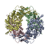
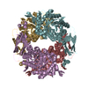
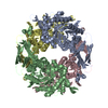
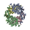
- PDB-1px8: Crystal structure of beta-D-xylosidase from Thermoanaerobacterium... -
+ Open data

- Basic information
| Entry | Database: PDB / ID: 1px8 | |||||||||
|---|---|---|---|---|---|---|---|---|---|---|
| Title | Crystal structure of beta-D-xylosidase from Thermoanaerobacterium saccharolyticum, a family 39 glycoside hydrolase | |||||||||
 Components Components | Beta-xylosidase | |||||||||
 Keywords Keywords | HYDROLASE / family 39 glycoside hydrolase / xylosidase / xylan / xylose / covalent glycosyl-enzyme intermediate | |||||||||
| Function / homology |  Function and homology information Function and homology informationxylan 1,4-beta-xylosidase / xylan 1,4-beta-xylosidase activity / xylan catabolic process Similarity search - Function | |||||||||
| Biological species |  Thermoanaerobacterium saccharolyticum (bacteria) Thermoanaerobacterium saccharolyticum (bacteria) | |||||||||
| Method |  X-RAY DIFFRACTION / SYNCHROTRON / MAD / Resolution: 2.4 Å X-RAY DIFFRACTION / SYNCHROTRON / MAD / Resolution: 2.4 Å | |||||||||
 Authors Authors | Yang, J.K. / Yoon, H.J. / Ahn, H.J. / Il Lee, B. / Pedelacq, J.D. / Liong, E.C. / Berendzen, J. / Laivenieks, M. / Vieille, C. / Zeikus, G.J. ...Yang, J.K. / Yoon, H.J. / Ahn, H.J. / Il Lee, B. / Pedelacq, J.D. / Liong, E.C. / Berendzen, J. / Laivenieks, M. / Vieille, C. / Zeikus, G.J. / Vocadlo, D.J. / Withers, S.G. / Suh, S.W. | |||||||||
 Citation Citation | Journal: J.Mol.Biol. / Year: 2004 Title: Crystal structure of beta-D-xylosidase from Thermoanaerobacterium saccharolyticum, a family 39 glycoside hydrolase. Authors: Yang, J.K. / Yoon, H.J. / Ahn, H.J. / Lee, B.I. / Pedelacq, J.D. / Liong, E.C. / Berendzen, J. / Laivenieks, M. / Vieille, C. / Zeikus, G.J. / Vocadlo, D.J. / Withers, S.G. / Suh, S.W. | |||||||||
| History |
|
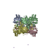
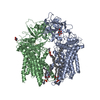
- Structure visualization
| Structure viewer | Molecule: MolmilJmol/JSmol |
|---|
- Downloads & links
-Download
| PDBx/mmCIF format | 1px8.cif.gz | 218.3 KB | Display | PDBx/mmCIF format |
|---|---|---|---|---|
| PDB format | pdb1px8.ent.gz | 175.1 KB | Display | PDB format |
| PDBx/mmJSON format | 1px8.json.gz | Tree view | PDBx/mmJSON format | |
| Others |  Other downloads Other downloads |
-Validation report
| Arichive directory | https://data.pdbj.org/pub/pdb/validation_reports/px/1px8ftp://data.pdbj.org/pub/pdb/validation_reports/px/1px8 | HTTPS FTP |
|---|
-Related structure data

-Links
 PDBj
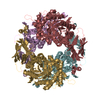
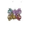
PDBj- Assembly
| Deposited unit |
| ||||||||
|---|---|---|---|---|---|---|---|---|---|
| 1 |
| ||||||||
| Unit cell |
|
-Components
| #1: Protein | Mass: 58739.180 Da / Num. of mol.: 2 Source method: isolated from a genetically manipulated source Source: (gene. exp.) Thermoanaerobacterium saccharolyticum (bacteria)Plasmid: pXHP3 / Production host: #2: Sugar | 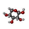  Type: D-saccharide, beta linking / Mass: 150.130 Da / Num. of mol.: 2 Type: D-saccharide, beta linking / Mass: 150.130 Da / Num. of mol.: 2Source method: isolated from a genetically manipulated source Formula: C5H10O5 #3: Water | ChemComp-HOH / |  Mass: 18.015 Da / Num. of mol.: 327 / Source method: isolated from a natural source / Formula: H2O Mass: 18.015 Da / Num. of mol.: 327 / Source method: isolated from a natural source / Formula: H2O |
|---|
-Experimental details
-Experiment
| Experiment | Method: X-RAY DIFFRACTION / Number of used crystals: 1 |
|---|
- Sample preparation
| Crystal | Density Matthews: 2.14 Å3/Da / Density % sol: 42.07 % | ||||||||||||||||||||||||||||||||||||
|---|---|---|---|---|---|---|---|---|---|---|---|---|---|---|---|---|---|---|---|---|---|---|---|---|---|---|---|---|---|---|---|---|---|---|---|---|---|
| Crystal grow | Temperature: 295 K / Method: vapor diffusion, hanging drop / pH: 7 Details: PEG 2000 MME, Tris-HCl, dithiothreitol, pH 7.0, VAPOR DIFFUSION, HANGING DROP, temperature 295K | ||||||||||||||||||||||||||||||||||||
| Crystal grow | *PLUS Temperature: 296 K / Method: vapor diffusion, hanging drop / Details: Yang, J.K., (2002) Acta Crystallogr., D58, 531. | ||||||||||||||||||||||||||||||||||||
| Components of the solutions | *PLUS
|
-Data collection
| Diffraction | Mean temperature: 100 K | |||||||||||||||
|---|---|---|---|---|---|---|---|---|---|---|---|---|---|---|---|---|
| Diffraction source | Source: SYNCHROTRON / Site: PAL/PLS  / Beamline: 6B / Wavelength: 0.9500, 0.9787, 0.9789, 1.0000 / Beamline: 6B / Wavelength: 0.9500, 0.9787, 0.9789, 1.0000 | |||||||||||||||
| Detector | Type: MACSCIENCE / Detector: IMAGE PLATE / Date: Oct 10, 1998 | |||||||||||||||
| Radiation | Protocol: MAD / Monochromatic (M) / Laue (L): M / Scattering type: x-ray | |||||||||||||||
| Radiation wavelength |
| |||||||||||||||
| Reflection | Resolution: 2.4→20 Å / Num. all: 37528 / Num. obs: 35679 / % possible obs: 89.1 % / Observed criterion σ(F): 0 / Observed criterion σ(I): 0 / Biso Wilson estimate: 12 Å2 / Rsym value: 0.066 / Net I/σ(I): 15.1 | |||||||||||||||
| Reflection shell | Resolution: 2.4→2.49 Å / Mean I/σ(I) obs: 2.3 / Rsym value: 0.247 / % possible all: 76.6 | |||||||||||||||
| Reflection | *PLUS Num. obs: 37528 / Num. measured all: 172649 / Rmerge(I) obs: 0.066 | |||||||||||||||
| Reflection shell | *PLUS % possible obs: 76.6 % / Rmerge(I) obs: 0.247 |
- Processing
| Software |
| ||||||||||||||||||||||||||||||||||||||||||||||||||||||||||||||||||||||||||||||||
|---|---|---|---|---|---|---|---|---|---|---|---|---|---|---|---|---|---|---|---|---|---|---|---|---|---|---|---|---|---|---|---|---|---|---|---|---|---|---|---|---|---|---|---|---|---|---|---|---|---|---|---|---|---|---|---|---|---|---|---|---|---|---|---|---|---|---|---|---|---|---|---|---|---|---|---|---|---|---|---|---|---|
| Refinement | Method to determine structure: MAD / Resolution: 2.4→19.91 Å / Rfactor Rfree error: 0.005 / Isotropic thermal model: RESTRAINED / Cross valid method: THROUGHOUT / σ(F): 2 / Stereochemistry target values: Engh & Huber
| ||||||||||||||||||||||||||||||||||||||||||||||||||||||||||||||||||||||||||||||||
| Solvent computation | Solvent model: FLAT MODEL / Bsol: 12.7696 Å2 / ksol: 0.28452 e/Å3 | ||||||||||||||||||||||||||||||||||||||||||||||||||||||||||||||||||||||||||||||||
| Displacement parameters | Biso mean: 37.9 Å2
| ||||||||||||||||||||||||||||||||||||||||||||||||||||||||||||||||||||||||||||||||
| Refine analyze | Luzzati coordinate error free: 0.4 Å / Luzzati sigma a free: 0.57 Å | ||||||||||||||||||||||||||||||||||||||||||||||||||||||||||||||||||||||||||||||||
| Refinement step | Cycle: LAST / Resolution: 2.4→19.91 Å
| ||||||||||||||||||||||||||||||||||||||||||||||||||||||||||||||||||||||||||||||||
| Refine LS restraints |
| ||||||||||||||||||||||||||||||||||||||||||||||||||||||||||||||||||||||||||||||||
| Refine LS restraints NCS | NCS model details: CONSTR | ||||||||||||||||||||||||||||||||||||||||||||||||||||||||||||||||||||||||||||||||
| LS refinement shell | Resolution: 2.4→2.55 Å / Rfactor Rfree error: 0.016 / Total num. of bins used: 6
| ||||||||||||||||||||||||||||||||||||||||||||||||||||||||||||||||||||||||||||||||
| Xplor file |
| ||||||||||||||||||||||||||||||||||||||||||||||||||||||||||||||||||||||||||||||||
| Refinement | *PLUS Highest resolution: 2.4 Å / Lowest resolution: 20 Å / % reflection Rfree: 10 % | ||||||||||||||||||||||||||||||||||||||||||||||||||||||||||||||||||||||||||||||||
| Solvent computation | *PLUS | ||||||||||||||||||||||||||||||||||||||||||||||||||||||||||||||||||||||||||||||||
| Displacement parameters | *PLUS | ||||||||||||||||||||||||||||||||||||||||||||||||||||||||||||||||||||||||||||||||
| Refine LS restraints | *PLUS
|
