SHEET DETERMINATION METHOD: DSSP THE SHEETS PRESENTED AS "AA" IN EACH CHAIN ON SHEET RECORDS BELOW ... SHEET DETERMINATION METHOD: DSSP THE SHEETS PRESENTED AS "AA" IN EACH CHAIN ON SHEET RECORDS BELOW IS ACTUALLY AN 9-STRANDED BARREL THIS IS REPRESENTED BY A 10-STRANDED SHEET IN WHICH THE FIRST AND LAST STRANDS ARE IDENTICAL. THE SHEETS PRESENTED AS "BA, CA" IN EACH CHAIN ON SHEET RECORDS BELOW ARE ACTUALLY 8-STRANDED BARRELS REPRESENTED BY 9-STRANDED SHEETS IN WHICH THE FIRST AND LAST STRANDS ARE IDENTICAL. THE SHEETS PRESENTED AS "DA, EA, FA, GA, HB " IN EACH CHAIN ON SHEET RECORDS BELOW ARE ACTUALLY 7-STRANDED BARRELS REPRESENTED BY 8-STRANDED SHEETS IN WHICH THE FIRST AND LAST STRANDS ARE IDENTICAL.
Component-ID: 1 / Beg auth comp-ID: VAL / Beg label comp-ID: VAL / End auth comp-ID: SER / End label comp-ID: SER / Refine code: 4 / Auth seq-ID: 4 - 502 / Label seq-ID: 4 - 502
Mass: 18.015 Da / Num. of mol.: 2889 / Source method: isolated from a natural source / Formula: H2O
Sequence details
THE PROTEIN CORRESPONDS TO THE UNIPROT ENTRY Q9ZFM2. THE SAMPLE USED IN THE EXPERIMENT HAD AN ...THE PROTEIN CORRESPONDS TO THE UNIPROT ENTRY Q9ZFM2. THE SAMPLE USED IN THE EXPERIMENT HAD AN INSERTION AT POSITION 445 IN THE PDB ENTRY AND THE RESIDUES 249-250 FROM THE UNIPROT ENTRY WERE DELETED. THE DBREF RECORD DESCRIBES THE DELETION AND INSERTION MUTATION. THE SEQADV RECORDS DESCRIBE THE CONFLICTS BETWEEN THE UNIPROT ENTRY Q9ZFM2 AND THE OBSERVED CRYSTAL STRUCTURE.
-
Experimental details
-
Experiment
Experiment
Method: X-RAY DIFFRACTION
-
Sample preparation
Crystal
Density Matthews: 2.62 Å3/Da / Density % sol: 53 %
Method to determine structure: OTHER / Resolution: 2.2→8 Å / Cor.coef. Fo:Fc: 0.952 / Cor.coef. Fo:Fc free: 0.909 / SU B: 5.186 / SU ML: 0.136 / Cross valid method: THROUGHOUT / ESU R: 0.257 / ESU R Free: 0.216 / Stereochemistry target values: MAXIMUM LIKELIHOOD / Details: HYDROGENS HAVE BEEN ADDED IN THE RIDING POSITIONS
Rfactor
Num. reflection
% reflection
Selection details
Rfree
0.242
11807
5 %
RANDOM
Rwork
0.174
-
-
-
obs
0.177
223893
100 %
-
Solvent computation
Ion probe radii: 0.8 Å / Shrinkage radii: 0.8 Å / VDW probe radii: 1.2 Å / Solvent model: MASK
 Movie
Movie Controller

 Yorodumi
 Open data
 Basic information
 Components
 Keywords
 Function and homology information
 GEOBACILLUS STEAROTHERMOPHILUS (bacteria)
 X-RAY DIFFRACTION /
  Authors
 Citation
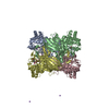
 Structure visualization
 Downloads & links
 Other downloads


 PDBj
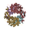
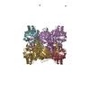
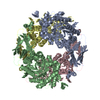
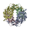
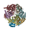
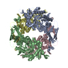
PDBj Assembly
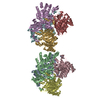
 Mass: 18.015 Da / Num. of mol.: 2889 / Source method: isolated from a natural source / Formula: H2O
 Sample preparation
 / Beamline: BM14 / Wavelength: 0.93
  Processing