Mass: 18.015 Da / Num. of mol.: 291 / Source method: isolated from a natural source / Formula: H2O
Compound details
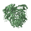
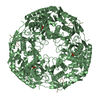
THIS IS A BIFUNCTIONAL ENZYME EC:2.7.7.8- POLYRIBONUCLEOTIDE NUCLEOTIDYLTRANSFERASE EC:2.7.6.5- ATP: ...THIS IS A BIFUNCTIONAL ENZYME EC:2.7.7.8- POLYRIBONUCLEOTIDE NUCLEOTIDYLTRANSFERASE EC:2.7.6.5- ATP:GTP 3'-PYROPHOSPHOTRANSFERASE
Has protein modification
Y
Sequence details
ARG A 31, SEQUENCING AMBIGUITY ILE A 156, SEQUENCING AMBIGUITY ILE A 210, SEQUENCING AMBIGUITY PHE ...ARG A 31, SEQUENCING AMBIGUITY ILE A 156, SEQUENCING AMBIGUITY ILE A 210, SEQUENCING AMBIGUITY PHE A 260, SEQUENCING AMBIGUITY LEU A 261, SEQUENCING AMBIGUITY ASP A 262, SEQUENCING AMBIGUITY TYR A 263, SEQUENCING AMBIGUITY GLN A 264, SEQUENCING AMBIGUITY ASP A 265, SEQUENCING AMBIGUITY VAL A 267, SEQUENCING AMBIGUITY LEU A 268, SEQUENCING AMBIGUITY GLU A 269, SEQUENCING AMBIGUITY ALA A 323, SEQUENCING AMBIGUITY LEU A 324, SEQUENCING AMBIGUITY THR A 325, SEQUENCING AMBIGUITY LYS A 326, SEQUENCING AMBIGUITY LEU A 328, SEQUENCING AMBIGUITY VAL A 329, SEQUENCING AMBIGUITY ARG A 330, SEQUENCING AMBIGUITY ALA A 335, SEQUENCING AMBIGUITY TYR A 409, SEQUENCING AMBIGUITY
-
Experimental details
-
Experiment
Experiment
Method: X-RAY DIFFRACTION / Number of used crystals: 1
-
Sample preparation
Crystal
Density Matthews: 3.6 Å3/Da / Density % sol: 60 % Description: DATA STATISTICS ARE FOR LOW ENERGY REMOTE DATASET. INITIAL SE SUBSTRUCTURE MODEL FROM SHELX-90 SEARCH OF REVISE PATTERSON MAP.
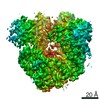
Resolution: 2.6→38.99 Å / Num. obs: 36704 / % possible obs: 99.6 % / Redundancy: 4.6 % / Biso Wilson estimate: 53.6 Å2 / Rsym value: 0.086 / Net I/σ(I): 5.3
Reflection shell
Resolution: 2.6→2.74 Å / Redundancy: 4.2 % / Mean I/σ(I) obs: 1.1 / Rsym value: 0.59 / % possible all: 98.1
-
Processing
Software
Name
Version
Classification
DENZO
datareduction
SCALA
datascaling
CNS
0.9
phasing
SHARP
phasing
CNS
0.9
refinement
Refinement
Method to determine structure: MAD / Resolution: 2.6→38.99 Å / Rfactor Rfree error: 0.005 / Data cutoff high absF: 9971455.35 / Isotropic thermal model: RESTRAINED / Cross valid method: THROUGHOUT / σ(F): 0 Details: REFINEMENT TARGET (MLHL) INCLUDED WEIGHTED EXPERIMENTAL PHASE DISTRIBUTION ESTIMATES FROM SHARP POOR DENSITY FOR RESIDUES 605 - 614 AND 623 - 632 WAS INTERPRETED FROM MODEL OF HOMOLOGOUS ...Details: REFINEMENT TARGET (MLHL) INCLUDED WEIGHTED EXPERIMENTAL PHASE DISTRIBUTION ESTIMATES FROM SHARP POOR DENSITY FOR RESIDUES 605 - 614 AND 623 - 632 WAS INTERPRETED FROM MODEL OF HOMOLOGOUS DOMAIN PDB 1VIH POSITIONED BY HAND IN DENSITY. MODEL HERE IS POLYALA (EXCEPT GLY AND PRO WHERE EXPECTED FROM SEQUENCE) WITH B-FACTOR SET TO 100.00 AND SUBJECT TO POSITIONAL REFINEMENT ONLY. THE C- TERMINAL RESIDUE WAS NOT SEEN IN DENSITY MAPS
In the structure databanks used in Yorodumi, some data are registered as the other names, "COVID-19 virus" and "2019-nCoV". Here are the details of the virus and the list of structure data.
Jan 31, 2019. EMDB accession codes are about to change! (news from PDBe EMDB page)
EMDB accession codes are about to change! (news from PDBe EMDB page)
The allocation of 4 digits for EMDB accession codes will soon come to an end. Whilst these codes will remain in use, new EMDB accession codes will include an additional digit and will expand incrementally as the available range of codes is exhausted. The current 4-digit format prefixed with “EMD-” (i.e. EMD-XXXX) will advance to a 5-digit format (i.e. EMD-XXXXX), and so on. It is currently estimated that the 4-digit codes will be depleted around Spring 2019, at which point the 5-digit format will come into force.
The EM Navigator/Yorodumi systems omit the EMD- prefix.
Related info.:Q: What is EMD? / ID/Accession-code notation in Yorodumi/EM Navigator
Yorodumi is a browser for structure data from EMDB, PDB, SASBDB, etc.
This page is also the successor to EM Navigator detail page, and also detail information page/front-end page for Omokage search.
The word "yorodu" (or yorozu) is an old Japanese word meaning "ten thousand". "mi" (miru) is to see.
Related info.:EMDB / PDB / SASBDB / Comparison of 3 databanks / Yorodumi Search / Aug 31, 2016. New EM Navigator & Yorodumi / Yorodumi Papers / Jmol/JSmol / Function and homology information / Changes in new EM Navigator and Yorodumi
 Movie
Movie Controller


 Open data
 Basic information
 Components
 Keywords
 Function and homology information
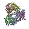
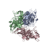
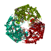
 STREPTOMYCES ANTIBIOTICUS (bacteria)
 X-RAY DIFFRACTION /
  Authors
 Citation
 Structure visualization
 Downloads & links
 Other downloads


 PDBj
PDBj


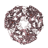
 Assembly

 Mass: 96.063 Da / Num. of mol.: 9 / Source method: obtained synthetically / Formula: SO4
 Mass: 18.015 Da / Num. of mol.: 291 / Source method: isolated from a natural source / Formula: H2O
 Sample preparation
 / Beamline: BW7A / Wavelength: 0.9740, 0.9791, 0.98
 Processing