 Movie
Movie Controller

[English] 日本語
 Yorodumi
- PDB-6bap: Stigmatella aurantiaca bacterial phytochrome PAS-GAF-PHY, T289H mutant -
+ Open data

- Basic information
| Entry | Database: PDB / ID: 6bap | |||||||||||||||
|---|---|---|---|---|---|---|---|---|---|---|---|---|---|---|---|---|
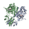
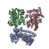
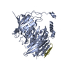
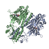
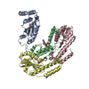
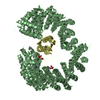
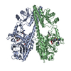
| Title | Stigmatella aurantiaca bacterial phytochrome PAS-GAF-PHY, T289H mutant | |||||||||||||||
 Components Components | Photoreceptor-histidine kinase BphP | |||||||||||||||
 Keywords Keywords | SIGNALING PROTEIN / phytochrome / photosensory core module / T289H mutant | |||||||||||||||
| Function / homology |  Function and homology information Function and homology informationosmosensory signaling via phosphorelay pathway / detection of visible light / phosphorelay response regulator activity / phosphorelay sensor kinase activity / histidine kinase / photoreceptor activity / protein kinase activator activity / regulation of DNA-templated transcription Similarity search - Function | |||||||||||||||
| Biological species |  Stigmatella aurantiaca DW4/3-1 (bacteria) Stigmatella aurantiaca DW4/3-1 (bacteria) | |||||||||||||||
| Method |  X-RAY DIFFRACTION / SYNCHROTRON / MOLECULAR REPLACEMENT / Resolution: 2.65 Å X-RAY DIFFRACTION / SYNCHROTRON / MOLECULAR REPLACEMENT / Resolution: 2.65 Å | |||||||||||||||
 Authors Authors | Schmidt, M. / Stojkovic, E. | |||||||||||||||
| Funding support |  United States, 4items United States, 4items
| |||||||||||||||
 Citation Citation | Journal: IUCrJ / Year: 2018 Title: Structural basis for light control of cell development revealed by crystal structures of a myxobacterial phytochrome. Authors: Woitowich, N.C. / Halavaty, A.S. / Waltz, P. / Kupitz, C. / Valera, J. / Tracy, G. / Gallagher, K.D. / Claesson, E. / Nakane, T. / Pandey, S. / Nelson, G. / Tanaka, R. / Nango, E. / ...Authors: Woitowich, N.C. / Halavaty, A.S. / Waltz, P. / Kupitz, C. / Valera, J. / Tracy, G. / Gallagher, K.D. / Claesson, E. / Nakane, T. / Pandey, S. / Nelson, G. / Tanaka, R. / Nango, E. / Mizohata, E. / Owada, S. / Tono, K. / Joti, Y. / Nugent, A.C. / Patel, H. / Mapara, A. / Hopkins, J. / Duong, P. / Bizhga, D. / Kovaleva, S.E. / St Peter, R. / Hernandez, C.N. / Ozarowski, W.B. / Roy-Chowdhuri, S. / Yang, J.H. / Edlund, P. / Takala, H. / Ihalainen, J. / Brayshaw, J. / Norwood, T. / Poudyal, I. / Fromme, P. / Spence, J.C.H. / Moffat, K. / Westenhoff, S. / Schmidt, M. / Stojkovic, E.A. | |||||||||||||||
| History |
|
- Structure visualization
| Structure viewer | Molecule: MolmilJmol/JSmol |
|---|
- Downloads & links
-Download
| PDBx/mmCIF format | 6bap.cif.gz | 312.1 KB | Display | PDBx/mmCIF format |
|---|---|---|---|---|
| PDB format | pdb6bap.ent.gz | 251.6 KB | Display | PDB format |
| PDBx/mmJSON format | 6bap.json.gz | Tree view | PDBx/mmJSON format | |
| Others |  Other downloads Other downloads |
-Validation report
| Arichive directory | https://data.pdbj.org/pub/pdb/validation_reports/ba/6bapftp://data.pdbj.org/pub/pdb/validation_reports/ba/6bap | HTTPS FTP |
|---|
-Related structure data
| Related structure data |  6bafC  6bakC  6baoSC  6bayC S: Starting model for refinement C: citing same article ( |
|---|---|
| Similar structure data |

-Links
 PDBj
PDBj


- Assembly
| Deposited unit | 
| ||||||||
|---|---|---|---|---|---|---|---|---|---|
| 1 | 
| ||||||||
| 2 | 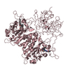
| ||||||||
| Unit cell |
|
-Components
| #1: Protein | Mass: 57548.180 Da / Num. of mol.: 3 / Fragment: PAS-GAF-PHY (UNP residues 1-515) / Mutation: T289H Source method: isolated from a genetically manipulated source Source: (gene. exp.) Stigmatella aurantiaca DW4/3-1 (bacteria)Strain: DW4/3-1 / Gene: STAUR_8015, STIAU_3396 / Production host: #2: Chemical |   Mass: 584.662 Da / Num. of mol.: 3 Mass: 584.662 Da / Num. of mol.: 3Source method: isolated from a genetically manipulated source Formula: C33H36N4O6 #3: Water | ChemComp-HOH / |  Mass: 18.015 Da / Num. of mol.: 124 / Source method: isolated from a natural source / Formula: H2O Mass: 18.015 Da / Num. of mol.: 124 / Source method: isolated from a natural source / Formula: H2OHas protein modification | Y | |
|---|
-Experimental details
-Experiment
| Experiment | Method: X-RAY DIFFRACTION / Number of used crystals: 1 |
|---|
- Sample preparation
| Crystal | Density Matthews: 2.82 Å3/Da / Density % sol: 56.33 % |
|---|---|
| Crystal grow | Temperature: 289 K / Method: vapor diffusion, hanging drop / pH: 6.2 Details: 11 mg/mL protein in 0.1 M MES, pH 6.2, 8-5% w/v PEG20000, 5.2% v/v acetonitrile |
-Data collection
| Diffraction | Mean temperature: 100 K |
|---|---|
| Diffraction source | Source: SYNCHROTRON / Site: APS / Beamline: 19-ID / Wavelength: 0.987 Å |
| Detector | Type: ADSC QUANTUM 315r / Detector: CCD / Date: Nov 23, 2013 |
| Radiation | Monochromator: double crystal Si(111) / Protocol: SINGLE WAVELENGTH / Monochromatic (M) / Laue (L): M / Scattering type: x-ray |
| Radiation wavelength | Wavelength: 0.987 Å / Relative weight: 1 |
| Reflection | Resolution: 2.65→42.6 Å / Num. obs: 56756 / % possible obs: 99.1 % / Redundancy: 3.5 % / CC1/2: 0.997 / Rpim(I) all: 0.041 / Rrim(I) all: 0.084 / Rsym value: 0.073 / Net I/σ(I): 7.9 |
| Reflection shell | Resolution: 2.65→2.72 Å / Redundancy: 3.3 % / Mean I/σ(I) obs: 0.8 / Num. unique obs: 4109 / CC1/2: 0.364 / Rpim(I) all: 0.553 / Rrim(I) all: 1.081 / Rsym value: 0.919 / % possible all: 99.1 |
- Processing
| Software |
| |||||||||||||||||||||||||||||||||||||||||||||||||||||||||||||||||||||||||||||||||||||||||||||||||||||||||||||||||||||||||||||||||||||||||||||||||||
|---|---|---|---|---|---|---|---|---|---|---|---|---|---|---|---|---|---|---|---|---|---|---|---|---|---|---|---|---|---|---|---|---|---|---|---|---|---|---|---|---|---|---|---|---|---|---|---|---|---|---|---|---|---|---|---|---|---|---|---|---|---|---|---|---|---|---|---|---|---|---|---|---|---|---|---|---|---|---|---|---|---|---|---|---|---|---|---|---|---|---|---|---|---|---|---|---|---|---|---|---|---|---|---|---|---|---|---|---|---|---|---|---|---|---|---|---|---|---|---|---|---|---|---|---|---|---|---|---|---|---|---|---|---|---|---|---|---|---|---|---|---|---|---|---|---|---|---|---|
| Refinement | Method to determine structure: MOLECULAR REPLACEMENT Starting model: PDB entry 6BAO Resolution: 2.65→40.284 Å / SU ML: 0.52 / Cross valid method: FREE R-VALUE / σ(F): 1.34 / Phase error: 32.78
| |||||||||||||||||||||||||||||||||||||||||||||||||||||||||||||||||||||||||||||||||||||||||||||||||||||||||||||||||||||||||||||||||||||||||||||||||||
| Solvent computation | Shrinkage radii: 0.9 Å / VDW probe radii: 1.11 Å | |||||||||||||||||||||||||||||||||||||||||||||||||||||||||||||||||||||||||||||||||||||||||||||||||||||||||||||||||||||||||||||||||||||||||||||||||||
| Refinement step | Cycle: LAST / Resolution: 2.65→40.284 Å
| |||||||||||||||||||||||||||||||||||||||||||||||||||||||||||||||||||||||||||||||||||||||||||||||||||||||||||||||||||||||||||||||||||||||||||||||||||
| Refine LS restraints |
| |||||||||||||||||||||||||||||||||||||||||||||||||||||||||||||||||||||||||||||||||||||||||||||||||||||||||||||||||||||||||||||||||||||||||||||||||||
| LS refinement shell |
|
