 Movie
Movie Controller

[English] 日本語
 Yorodumi
- PDB-2vou: Structure of 2,6-dihydroxypyridine-3-hydroxylase from Arthrobacte... -
+ Open data

- Basic information
| Entry | Database: PDB / ID: 2vou | ||||||
|---|---|---|---|---|---|---|---|
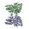
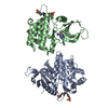
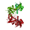
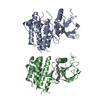
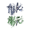
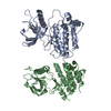
| Title | Structure of 2,6-dihydroxypyridine-3-hydroxylase from Arthrobacter nicotinovorans | ||||||
 Components Components | 2,6-DIHYDROXYPYRIDINE HYDROXYLASE | ||||||
 Keywords Keywords | OXIDOREDUCTASE / AROMATIC HYDROXYLASE / NICOTINE DEGRADATION / FLAVIN MONO-OXYGENASE / FAD-DEPENDENT HYDROXYLASE | ||||||
| Function / homology |  Function and homology information Function and homology information2,6-dihydroxypyridine 3-monooxygenase / 2,6-dihydroxypyridine 3-monooxygenase activity / nicotine catabolic process / FAD binding / flavin adenine dinucleotide binding / protein homodimerization activity Similarity search - Function | ||||||
| Biological species |  ARTHROBACTER NICOTINOVORANS (bacteria) ARTHROBACTER NICOTINOVORANS (bacteria) | ||||||
| Method |  X-RAY DIFFRACTION / SYNCHROTRON / MAD / Resolution: 2.6 Å X-RAY DIFFRACTION / SYNCHROTRON / MAD / Resolution: 2.6 Å | ||||||
 Authors Authors | Treiber, N. / Schulz, G.E. | ||||||
 Citation Citation | Journal: J.Mol.Biol. / Year: 2008 Title: Structure of 2,6-Dihydroxypyridine 3-Hydroxylase from a Nicotine-Degrading Pathway. Authors: Treiber, N. / Schulz, G.E. | ||||||
| History |
|
- Structure visualization
| Structure viewer | Molecule: MolmilJmol/JSmol |
|---|
- Downloads & links
-Download
| PDBx/mmCIF format | 2vou.cif.gz | 239.7 KB | Display | PDBx/mmCIF format |
|---|---|---|---|---|
| PDB format | pdb2vou.ent.gz | 194.7 KB | Display | PDB format |
| PDBx/mmJSON format | 2vou.json.gz | Tree view | PDBx/mmJSON format | |
| Others |  Other downloads Other downloads |
-Validation report
| Arichive directory | https://data.pdbj.org/pub/pdb/validation_reports/vo/2vouftp://data.pdbj.org/pub/pdb/validation_reports/vo/2vou | HTTPS FTP |
|---|
-Related structure data
| Similar structure data |
|---|


-Links
 PDBj
PDBj- Assembly
| Deposited unit | 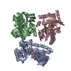
| ||||||||
|---|---|---|---|---|---|---|---|---|---|
| 1 | 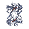
| ||||||||
| 2 | 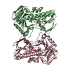
| ||||||||
| Unit cell |
| ||||||||
| Noncrystallographic symmetry (NCS) | NCS oper: (Code: given Matrix: (-0.5075, -0.79698, 0.32751), Vector: |
-Components
| #1: Protein | Mass: 43429.449 Da / Num. of mol.: 3 / Mutation: YES Source method: isolated from a genetically manipulated source Source: (gene. exp.) ARTHROBACTER NICOTINOVORANS (bacteria) / Production host: References: UniProt: Q93NG3, 2,6-dihydroxypyridine 3-monooxygenase #2: Chemical |   Mass: 785.550 Da / Num. of mol.: 3 / Source method: obtained synthetically / Formula: C27H33N9O15P2 / Comment: FAD*YM Mass: 785.550 Da / Num. of mol.: 3 / Source method: obtained synthetically / Formula: C27H33N9O15P2 / Comment: FAD*YM#3: Chemical |   Mass: 92.094 Da / Num. of mol.: 3 / Source method: obtained synthetically / Formula: C3H8O3 Mass: 92.094 Da / Num. of mol.: 3 / Source method: obtained synthetically / Formula: C3H8O3#4: Chemical |   Mass: 59.044 Da / Num. of mol.: 3 / Source method: obtained synthetically / Formula: C2H3O2 Mass: 59.044 Da / Num. of mol.: 3 / Source method: obtained synthetically / Formula: C2H3O2#5: Water | ChemComp-HOH / |  Mass: 18.015 Da / Num. of mol.: 387 / Source method: isolated from a natural source / Formula: H2O Mass: 18.015 Da / Num. of mol.: 387 / Source method: isolated from a natural source / Formula: H2OCompound details | ENGINEERED RESIDUE IN CHAIN A, CYS 323 TO SER ENGINEERED RESIDUE IN CHAIN B, CYS 323 TO SER ...ENGINEERED | |
|---|
-Experimental details
-Experiment
| Experiment | Method: X-RAY DIFFRACTION / Number of used crystals: 1 |
|---|
- Sample preparation
| Crystal | Density Matthews: 4.02 Å3/Da / Density % sol: 69 % / Description: NONE |
|---|---|
| Crystal grow | pH: 6.6 Details: 0.1 M SODIUM CACODYLATE PH 6.6, 15% (W/V) PEG 8000, 200 MM MAGNESIUM ACETATE, 20% (V/V) GLYCEROL |
-Data collection
| Diffraction | Mean temperature: 100 K |
|---|---|
| Diffraction source | Source: SYNCHROTRON / Site: SLS  / Beamline: X10SA / Wavelength: 1.0008 / Beamline: X10SA / Wavelength: 1.0008 |
| Detector | Type: MARRESEARCH / Detector: CCD / Date: Sep 5, 2006 |
| Radiation | Protocol: SINGLE WAVELENGTH / Monochromatic (M) / Laue (L): M / Scattering type: x-ray |
| Radiation wavelength | Wavelength: 1.0008 Å / Relative weight: 1 |
| Reflection | Resolution: 2.6→20 Å / Num. obs: 64088 / % possible obs: 99.7 % / Observed criterion σ(I): 2.4 / Redundancy: 4.9 % / Biso Wilson estimate: 66.3 Å2 / Rmerge(I) obs: 0.07 / Net I/σ(I): 15 |
| Reflection shell | Resolution: 2.6→2.8 Å / Rmerge(I) obs: 0.51 / Mean I/σ(I) obs: 2.4 / % possible all: 99.5 |
- Processing
| Software |
| ||||||||||||||||||||||||||||||||||||||||||||||||||||||||||||||||||||||||||||||||||||||||||||||||||||||||||||||||||||||||||||||||||||||||||||||||||||||||||||||||||||||||||||||||||||||
|---|---|---|---|---|---|---|---|---|---|---|---|---|---|---|---|---|---|---|---|---|---|---|---|---|---|---|---|---|---|---|---|---|---|---|---|---|---|---|---|---|---|---|---|---|---|---|---|---|---|---|---|---|---|---|---|---|---|---|---|---|---|---|---|---|---|---|---|---|---|---|---|---|---|---|---|---|---|---|---|---|---|---|---|---|---|---|---|---|---|---|---|---|---|---|---|---|---|---|---|---|---|---|---|---|---|---|---|---|---|---|---|---|---|---|---|---|---|---|---|---|---|---|---|---|---|---|---|---|---|---|---|---|---|---|---|---|---|---|---|---|---|---|---|---|---|---|---|---|---|---|---|---|---|---|---|---|---|---|---|---|---|---|---|---|---|---|---|---|---|---|---|---|---|---|---|---|---|---|---|---|---|---|---|
| Refinement | Method to determine structure: MAD Starting model: NONE Resolution: 2.6→27.92 Å / Cor.coef. Fo:Fc: 0.959 / Cor.coef. Fo:Fc free: 0.942 / SU B: 18.65 / SU ML: 0.202 / TLS residual ADP flag: LIKELY RESIDUAL / Cross valid method: THROUGHOUT / ESU R: 0.318 / ESU R Free: 0.232 / Stereochemistry target values: MAXIMUM LIKELIHOOD / Details: HYDROGENS HAVE BEEN ADDED IN THE RIDING POSITIONS.
| ||||||||||||||||||||||||||||||||||||||||||||||||||||||||||||||||||||||||||||||||||||||||||||||||||||||||||||||||||||||||||||||||||||||||||||||||||||||||||||||||||||||||||||||||||||||
| Solvent computation | Ion probe radii: 0.8 Å / Shrinkage radii: 0.8 Å / VDW probe radii: 1.4 Å / Solvent model: MASK | ||||||||||||||||||||||||||||||||||||||||||||||||||||||||||||||||||||||||||||||||||||||||||||||||||||||||||||||||||||||||||||||||||||||||||||||||||||||||||||||||||||||||||||||||||||||
| Displacement parameters | Biso mean: 67.04 Å2
| ||||||||||||||||||||||||||||||||||||||||||||||||||||||||||||||||||||||||||||||||||||||||||||||||||||||||||||||||||||||||||||||||||||||||||||||||||||||||||||||||||||||||||||||||||||||
| Refinement step | Cycle: LAST / Resolution: 2.6→27.92 Å
| ||||||||||||||||||||||||||||||||||||||||||||||||||||||||||||||||||||||||||||||||||||||||||||||||||||||||||||||||||||||||||||||||||||||||||||||||||||||||||||||||||||||||||||||||||||||
| Refine LS restraints |
|