 Movie
Movie Controller

[English] 日本語
 Yorodumi
- PDB-6r7r: Crystal structure of the glutamate transporter homologue GltTk in... -
+ Open data

- Basic information
| Entry | Database: PDB / ID: 6r7r | ||||||
|---|---|---|---|---|---|---|---|
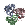
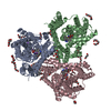
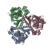
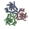
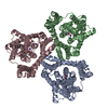
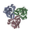
| Title | Crystal structure of the glutamate transporter homologue GltTk in complex with D-aspartate | ||||||
 Components Components | Proton/glutamate symporter, SDF family | ||||||
 Keywords Keywords | TRANSPORT PROTEIN / AMINO ACID TRANSPORTER / ASPARTATE TRANSPORT / GLUTAMATE TRANSPORTER HOMOLOGUE / MEMBRANE PROTEIN | ||||||
| Function / homology |  Function and homology information Function and homology informationdicarboxylic acid transport / symporter activity / membrane / plasma membrane Similarity search - Function | ||||||
| Biological species |   Thermococcus kodakarensis (archaea) Thermococcus kodakarensis (archaea) | ||||||
| Method |  X-RAY DIFFRACTION / SYNCHROTRON / MOLECULAR REPLACEMENT / Resolution: 2.8 Å X-RAY DIFFRACTION / SYNCHROTRON / MOLECULAR REPLACEMENT / Resolution: 2.8 Å | ||||||
 Authors Authors | Arkhipova, V. / Guskov, A. / Slotboom, D.J. | ||||||
| Funding support |  Netherlands, 1items Netherlands, 1items
| ||||||
 Citation Citation | Journal: Elife / Year: 2019 Title: Binding and transport of D-aspartate by the glutamate transporter homolog Glt Tk . Authors: Arkhipova, V. / Trinco, G. / Ettema, T.W. / Jensen, S. / Slotboom, D.J. / Guskov, A. | ||||||
| History |
|
- Structure visualization
| Structure viewer | Molecule: MolmilJmol/JSmol |
|---|
- Downloads & links
-Download
| PDBx/mmCIF format | 6r7r.cif.gz | 487.5 KB | Display | PDBx/mmCIF format |
|---|---|---|---|---|
| PDB format | pdb6r7r.ent.gz | 410.6 KB | Display | PDB format |
| PDBx/mmJSON format | 6r7r.json.gz | Tree view | PDBx/mmJSON format | |
| Others |  Other downloads Other downloads |
-Validation report
| Arichive directory | https://data.pdbj.org/pub/pdb/validation_reports/r7/6r7rftp://data.pdbj.org/pub/pdb/validation_reports/r7/6r7r | HTTPS FTP |
|---|
-Related structure data
| Related structure data | 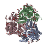 5e9sS S: Starting model for refinement |
|---|---|
| Similar structure data |


-Links
 PDBj
PDBj
- Assembly
| Deposited unit | 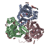
| ||||||||
|---|---|---|---|---|---|---|---|---|---|
| 1 |
| ||||||||
| Unit cell |
|
-Components
-Protein / Sugars , 2 types, 4 molecules ABC
| #1: Protein | Mass: 46685.363 Da / Num. of mol.: 3 Source method: isolated from a genetically manipulated source Source: (gene. exp.) Thermococcus kodakarensis (strain ATCC BAA-918 / JCM 12380 / KOD1) (archaea)Strain: ATCC BAA-918 / JCM 12380 / KOD1 / Gene: TK0986 / Production host:  #6: Sugar | ChemComp-DMU / |  Type: D-saccharide / Mass: 482.562 Da / Num. of mol.: 1 Type: D-saccharide / Mass: 482.562 Da / Num. of mol.: 1Source method: isolated from a genetically manipulated source Formula: C22H42O11 / Comment: detergent*YM |
|---|
-Non-polymers , 7 types, 31 molecules 


| #2: Chemical | ChemComp-NA /  Mass: 22.990 Da / Num. of mol.: 9 / Source method: obtained synthetically / Formula: Na Mass: 22.990 Da / Num. of mol.: 9 / Source method: obtained synthetically / Formula: Na#3: Chemical |  Type: D-peptide linking / Mass: 133.103 Da / Num. of mol.: 3 / Source method: obtained synthetically / Formula: C4H7NO4 / Feature type: SUBJECT OF INVESTIGATION Type: D-peptide linking / Mass: 133.103 Da / Num. of mol.: 3 / Source method: obtained synthetically / Formula: C4H7NO4 / Feature type: SUBJECT OF INVESTIGATION#4: Chemical | ChemComp-PEG /  Mass: 106.120 Da / Num. of mol.: 12 / Source method: obtained synthetically / Formula: C4H10O3 Mass: 106.120 Da / Num. of mol.: 12 / Source method: obtained synthetically / Formula: C4H10O3#5: Chemical | ChemComp-PG4 / |  Mass: 194.226 Da / Num. of mol.: 1 / Source method: obtained synthetically / Formula: C8H18O5 / Comment: precipitant*YM Mass: 194.226 Da / Num. of mol.: 1 / Source method: obtained synthetically / Formula: C8H18O5 / Comment: precipitant*YM#7: Chemical |  Mass: 150.173 Da / Num. of mol.: 3 / Source method: obtained synthetically / Formula: C6H14O4 Mass: 150.173 Da / Num. of mol.: 3 / Source method: obtained synthetically / Formula: C6H14O4#8: Chemical |  Mass: 282.331 Da / Num. of mol.: 2 / Source method: obtained synthetically / Formula: C12H26O7 / Comment: precipitant*YM Mass: 282.331 Da / Num. of mol.: 2 / Source method: obtained synthetically / Formula: C12H26O7 / Comment: precipitant*YM#9: Chemical | ChemComp-1PE / |  Mass: 238.278 Da / Num. of mol.: 1 / Source method: obtained synthetically / Formula: C10H22O6 / Comment: precipitant*YM Mass: 238.278 Da / Num. of mol.: 1 / Source method: obtained synthetically / Formula: C10H22O6 / Comment: precipitant*YM |
|---|
-Experimental details
-Experiment
| Experiment | Method: X-RAY DIFFRACTION / Number of used crystals: 1 |
|---|
- Sample preparation
| Crystal | Density Matthews: 4.7 Å3/Da / Density % sol: 73.8 % |
|---|---|
| Crystal grow | Temperature: 277 K / Method: vapor diffusion, hanging drop / pH: 8 Details: 20% glycerol, 10% PEG 4000, 100 mM Tris/bicine, pH 8.0, 60 mM CaCl2, 60 mM MgCl2, 0.75% n-octyl-b-D-glucopyranoside |
-Data collection
| Diffraction | Mean temperature: 100 K / Serial crystal experiment: N |
|---|---|
| Diffraction source | Source: SYNCHROTRON / Site: ESRF  / Beamline: ID23-1 / Wavelength: 0.99187 Å / Beamline: ID23-1 / Wavelength: 0.99187 Å |
| Detector | Type: DECTRIS PILATUS 6M-F / Detector: PIXEL / Date: Feb 23, 2017 |
| Radiation | Protocol: SINGLE WAVELENGTH / Monochromatic (M) / Laue (L): M / Scattering type: x-ray |
| Radiation wavelength | Wavelength: 0.99187 Å / Relative weight: 1 |
| Reflection | Resolution: 2.8→48.057 Å / Num. obs: 61573 / % possible obs: 99.3 % / Redundancy: 4.89 % / CC1/2: 0.999 / Rmerge(I) obs: 0.111 / Net I/σ(I): 8.4 |
| Reflection shell | Resolution: 2.8→2.87 Å / Redundancy: 3.58 % / Rmerge(I) obs: 3.768 / Num. unique obs: 4459 / CC1/2: 0.117 / % possible all: 98.9 |
- Processing
| Software |
| |||||||||||||||||||||||||||||||||||||||||||||||||||||||||||||||||||||||||||||||||||||||||||||||||||||||||||||||||||||||||||||||||||||||||||||||||||||||||||||||||
|---|---|---|---|---|---|---|---|---|---|---|---|---|---|---|---|---|---|---|---|---|---|---|---|---|---|---|---|---|---|---|---|---|---|---|---|---|---|---|---|---|---|---|---|---|---|---|---|---|---|---|---|---|---|---|---|---|---|---|---|---|---|---|---|---|---|---|---|---|---|---|---|---|---|---|---|---|---|---|---|---|---|---|---|---|---|---|---|---|---|---|---|---|---|---|---|---|---|---|---|---|---|---|---|---|---|---|---|---|---|---|---|---|---|---|---|---|---|---|---|---|---|---|---|---|---|---|---|---|---|---|---|---|---|---|---|---|---|---|---|---|---|---|---|---|---|---|---|---|---|---|---|---|---|---|---|---|---|---|---|---|---|---|
| Refinement | Method to determine structure: MOLECULAR REPLACEMENT Starting model: 5E9S Resolution: 2.8→48.057 Å / Cross valid method: FREE R-VALUE / σ(F): 1.33
| |||||||||||||||||||||||||||||||||||||||||||||||||||||||||||||||||||||||||||||||||||||||||||||||||||||||||||||||||||||||||||||||||||||||||||||||||||||||||||||||||
| Solvent computation | Shrinkage radii: 0.9 Å / VDW probe radii: 1.11 Å | |||||||||||||||||||||||||||||||||||||||||||||||||||||||||||||||||||||||||||||||||||||||||||||||||||||||||||||||||||||||||||||||||||||||||||||||||||||||||||||||||
| Refinement step | Cycle: LAST / Resolution: 2.8→48.057 Å
| |||||||||||||||||||||||||||||||||||||||||||||||||||||||||||||||||||||||||||||||||||||||||||||||||||||||||||||||||||||||||||||||||||||||||||||||||||||||||||||||||
| Refine LS restraints |
| |||||||||||||||||||||||||||||||||||||||||||||||||||||||||||||||||||||||||||||||||||||||||||||||||||||||||||||||||||||||||||||||||||||||||||||||||||||||||||||||||
| LS refinement shell |
|
