 Movie
Movie Controller

[English] 日本語
 Yorodumi
- PDB-6adc: Crystal structure of the E148A mutant CLC-ec1 in the presence of ... -
+ Open data

- Basic information
| Entry | Database: PDB / ID: 6adc | ||||||
|---|---|---|---|---|---|---|---|
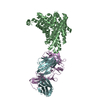
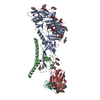
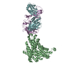
| Title | Crystal structure of the E148A mutant CLC-ec1 in the presence of 50mM bromoacetate | ||||||
 Components Components |
| ||||||
 Keywords Keywords | TRANSPORT PROTEIN / CLC Cl-/H+ antiporter / intermediate structure / external glutamate | ||||||
| Function / homology |  Function and homology information Function and homology informationcellular stress response to acidic pH / chloride:proton antiporter activity / voltage-gated chloride channel activity / proton transmembrane transport / chloride transmembrane transport / identical protein binding / plasma membrane Similarity search - Function | ||||||
| Biological species |   | ||||||
| Method |  X-RAY DIFFRACTION / SYNCHROTRON / MOLECULAR REPLACEMENT / molecular replacement / Resolution: 3.055 Å X-RAY DIFFRACTION / SYNCHROTRON / MOLECULAR REPLACEMENT / molecular replacement / Resolution: 3.055 Å | ||||||
 Authors Authors | Lim, H.-H. / Park, K. | ||||||
| Funding support |  Korea, Republic Of, 1items Korea, Republic Of, 1items
| ||||||
 Citation Citation | Journal: Proc.Natl.Acad.Sci.USA / Year: 2019 Title: Mutation of external glutamate residue reveals a new intermediate transport state and anion binding site in a CLC Cl-/H+antiporter. Authors: Park, K. / Lee, B.C. / Lim, H.H. | ||||||
| History |
|
- Structure visualization
| Structure viewer | Molecule: MolmilJmol/JSmol |
|---|
- Downloads & links
-Download
| PDBx/mmCIF format | 6adc.cif.gz | 336.1 KB | Display | PDBx/mmCIF format |
|---|---|---|---|---|
| PDB format | pdb6adc.ent.gz | 270.5 KB | Display | PDB format |
| PDBx/mmJSON format | 6adc.json.gz | Tree view | PDBx/mmJSON format | |
| Others |  Other downloads Other downloads |
-Validation report
| Arichive directory | https://data.pdbj.org/pub/pdb/validation_reports/ad/6adcftp://data.pdbj.org/pub/pdb/validation_reports/ad/6adc | HTTPS FTP |
|---|
-Related structure data
| Related structure data | 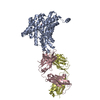 6ad7C 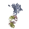 6ad8C 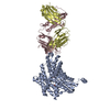 6adaC 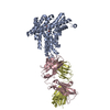 6adbC  6k5aC  6k5dC  6k5fC  6k5iC 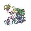 4eneS S: Starting model for refinement C: citing same article ( |
|---|---|
| Similar structure data |


-Links
 PDBj
PDBj

- Assembly
| Deposited unit | 
| ||||||||
|---|---|---|---|---|---|---|---|---|---|
| 1 | 
| ||||||||
| 2 | 
| ||||||||
| Unit cell |
|
-Components
| #1: Protein | Mass: 47042.754 Da / Num. of mol.: 2 / Mutation: E148A Source method: isolated from a genetically manipulated source Details: SF file contains Friedel pairs. Source: (gene. exp.) Strain: K12 / Gene: clcA, eriC, yadQ, b0155, JW5012 / Production host: #2: Antibody | Mass: 23823.031 Da / Num. of mol.: 2 / Source method: isolated from a natural source / Source: (natural) #3: Antibody | Mass: 23088.443 Da / Num. of mol.: 2 / Source method: isolated from a natural source / Source: (natural) #4: Chemical |   Mass: 138.948 Da / Num. of mol.: 2 / Source method: obtained synthetically / Formula: C2H3BrO2 Mass: 138.948 Da / Num. of mol.: 2 / Source method: obtained synthetically / Formula: C2H3BrO2Has protein modification | Y | |
|---|
-Experimental details
-Experiment
| Experiment | Method: X-RAY DIFFRACTION / Number of used crystals: 1 |
|---|
- Sample preparation
| Crystal | Density Matthews: 3.82 Å3/Da / Density % sol: 67.8 % |
|---|---|
| Crystal grow | Temperature: 295 K / Method: vapor diffusion, hanging drop / pH: 9 Details: PEG400 24% (w/v), 100mM glycine, 20 mM Na/K tartrate, 50 mM bromoacetate |
-Data collection
| Diffraction | Mean temperature: 80 K |
|---|---|
| Diffraction source | Source: SYNCHROTRON / Site: PAL/PLS / Beamline: 5C (4A) / Wavelength: 0.919 Å |
| Detector | Type: ADSC QUANTUM 315r / Detector: CCD / Date: Jun 12, 2018 |
| Radiation | Protocol: SINGLE WAVELENGTH / Monochromatic (M) / Laue (L): M / Scattering type: x-ray |
| Radiation wavelength | Wavelength: 0.919 Å / Relative weight: 1 |
| Reflection | Resolution: 3.055→49.9 Å / Num. obs: 102941 / % possible obs: 99.4 % / Redundancy: 7.5 % / Biso Wilson estimate: 102.39 Å2 / Rmerge(I) obs: 0.15 / Net I/σ(I): 32.1 |
| Reflection shell | Resolution: 3.05→3.16 Å / Redundancy: 7.4 % / Rmerge(I) obs: 1.33 / Mean I/σ(I) obs: 1.6 / Num. unique obs: 10254 / % possible all: 99.3 |
-Phasing
| Phasing | Method: molecular replacement |
|---|
- Processing
| Software |
| |||||||||||||||||||||||||||||||||||||||||||||||||||||||||||||||||||||||||||||||||||||||||||||||||||||||||||||||||||||||||||||||||||||||||||||||||||||||||||||||||||||||||||||||||||||||||||||||||||||||||||||||||||||||||
|---|---|---|---|---|---|---|---|---|---|---|---|---|---|---|---|---|---|---|---|---|---|---|---|---|---|---|---|---|---|---|---|---|---|---|---|---|---|---|---|---|---|---|---|---|---|---|---|---|---|---|---|---|---|---|---|---|---|---|---|---|---|---|---|---|---|---|---|---|---|---|---|---|---|---|---|---|---|---|---|---|---|---|---|---|---|---|---|---|---|---|---|---|---|---|---|---|---|---|---|---|---|---|---|---|---|---|---|---|---|---|---|---|---|---|---|---|---|---|---|---|---|---|---|---|---|---|---|---|---|---|---|---|---|---|---|---|---|---|---|---|---|---|---|---|---|---|---|---|---|---|---|---|---|---|---|---|---|---|---|---|---|---|---|---|---|---|---|---|---|---|---|---|---|---|---|---|---|---|---|---|---|---|---|---|---|---|---|---|---|---|---|---|---|---|---|---|---|---|---|---|---|---|---|---|---|---|---|---|---|---|---|---|---|---|---|---|---|---|
| Refinement | Method to determine structure: MOLECULAR REPLACEMENT Starting model: 4ENE Resolution: 3.055→34.852 Å / SU ML: 0.5 / Cross valid method: THROUGHOUT / σ(F): 0.01 / Phase error: 33.79 / Stereochemistry target values: ML
| |||||||||||||||||||||||||||||||||||||||||||||||||||||||||||||||||||||||||||||||||||||||||||||||||||||||||||||||||||||||||||||||||||||||||||||||||||||||||||||||||||||||||||||||||||||||||||||||||||||||||||||||||||||||||
| Solvent computation | Shrinkage radii: 0.9 Å / VDW probe radii: 1.11 Å / Solvent model: FLAT BULK SOLVENT MODEL | |||||||||||||||||||||||||||||||||||||||||||||||||||||||||||||||||||||||||||||||||||||||||||||||||||||||||||||||||||||||||||||||||||||||||||||||||||||||||||||||||||||||||||||||||||||||||||||||||||||||||||||||||||||||||
| Refinement step | Cycle: LAST / Resolution: 3.055→34.852 Å
| |||||||||||||||||||||||||||||||||||||||||||||||||||||||||||||||||||||||||||||||||||||||||||||||||||||||||||||||||||||||||||||||||||||||||||||||||||||||||||||||||||||||||||||||||||||||||||||||||||||||||||||||||||||||||
| Refine LS restraints |
| |||||||||||||||||||||||||||||||||||||||||||||||||||||||||||||||||||||||||||||||||||||||||||||||||||||||||||||||||||||||||||||||||||||||||||||||||||||||||||||||||||||||||||||||||||||||||||||||||||||||||||||||||||||||||
| LS refinement shell |
|
