 Movie
Movie Controller

[English] 日本語
 Yorodumi
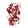
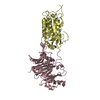
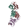
- PDB-5zbq: The Crystal Structure of human neuropeptide Y Y1 receptor with UR... -
+ Open data

- Basic information
| Entry | Database: PDB / ID: 5zbq | ||||||
|---|---|---|---|---|---|---|---|
| Title | The Crystal Structure of human neuropeptide Y Y1 receptor with UR-MK299 | ||||||
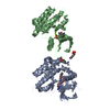
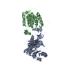
 Components Components | Neuropeptide Y receptor type 1,T4 Lysozyme | ||||||
 Keywords Keywords |  SIGNALING PROTEIN / G Protein-Coupled Receptor / Receptor Inhibitor / Complex structure SIGNALING PROTEIN / G Protein-Coupled Receptor / Receptor Inhibitor / Complex structure | ||||||
| Function / homology |  Function and homology information Function and homology informationpancreatic polypeptide receptor activity / peptide YY receptor activity / neuropeptide Y receptor activity / neuropeptide receptor activity / neuropeptide binding / feeding behavior / outflow tract morphogenesis / G protein-coupled receptor signaling pathway, coupled to cyclic nucleotide second messenger / regulation of multicellular organism growth / viral release from host cell by cytolysis ...pancreatic polypeptide receptor activity / peptide YY receptor activity / neuropeptide Y receptor activity / neuropeptide receptor activity / neuropeptide binding / feeding behavior / outflow tract morphogenesis / G protein-coupled receptor signaling pathway, coupled to cyclic nucleotide second messenger / regulation of multicellular organism growth / viral release from host cell by cytolysis / sensory perception of pain / adenylate cyclase-inhibiting G protein-coupled receptor signaling pathway / Peptide ligand-binding receptors / peptidoglycan catabolic process / locomotory behavior / regulation of blood pressure / glucose metabolic process / cell wall macromolecule catabolic process / lysozyme / lysozyme activity / G alpha (i) signalling events / host cell cytoplasm / defense response to bacterium / neuron projection / plasma membraneSimilarity search - Function | ||||||
| Biological species |  Homo sapiens (human) Homo sapiens (human) Enterobacteria phage RB55 (virus) Enterobacteria phage RB55 (virus) | ||||||
| Method | X-RAY DIFFRACTION / SYNCHROTRON / MOLECULAR REPLACEMENT / Resolution: 2.7 Å | ||||||
 Authors Authors | Yang, Z. / Han, S. / Zhao, Q. / Wu, B. | ||||||
 Citation Citation | Journal: Nature / Year: 2018 Title: Structural basis of ligand binding modes at the neuropeptide Y Y1receptor Authors: Yang, Z. / Han, S. / Keller, M. / Kaiser, A. / Bender, B.J. / Bosse, M. / Burkert, K. / Kogler, L.M. / Wifling, D. / Bernhardt, G. / Plank, N. / Littmann, T. / Schmidt, P. / Yi, C. / Li, B. ...Authors: Yang, Z. / Han, S. / Keller, M. / Kaiser, A. / Bender, B.J. / Bosse, M. / Burkert, K. / Kogler, L.M. / Wifling, D. / Bernhardt, G. / Plank, N. / Littmann, T. / Schmidt, P. / Yi, C. / Li, B. / Ye, S. / Zhang, R. / Xu, B. / Larhammar, D. / Stevens, R.C. / Huster, D. / Meiler, J. / Zhao, Q. / Beck-Sickinger, A.G. / Buschauer, A. / Wu, B. | ||||||
| History |
|
- Structure visualization
| Structure viewer | Molecule: MolmilJmol/JSmol |
|---|
- Downloads & links
-Download
| PDBx/mmCIF format | 5zbq.cif.gz | 159.5 KB | Display | PDBx/mmCIF format |
|---|---|---|---|---|
| PDB format | pdb5zbq.ent.gz | 117.9 KB | Display | PDB format |
| PDBx/mmJSON format | 5zbq.json.gz | Tree view | PDBx/mmJSON format | |
| Others |  Other downloads Other downloads |
-Validation report
| Arichive directory | https://data.pdbj.org/pub/pdb/validation_reports/zb/5zbqftp://data.pdbj.org/pub/pdb/validation_reports/zb/5zbq | HTTPS FTP |
|---|
-Related structure data
| Related structure data |  5zbhC 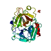 1c5pS  4grvS C: citing same article ( S: Starting model for refinement |
|---|---|
| Similar structure data |


-Links
 PDBj
PDBj


- Assembly
| Deposited unit | 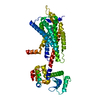
| ||||||||
|---|---|---|---|---|---|---|---|---|---|
| 1 |
| ||||||||
| Unit cell |
| ||||||||
| Details | AUTHORS STATE THAT THE BIOLOGICAL UNIT IS NUKNOWN |
-Components
| #1: Protein | Mass: 60595.223 Da / Num. of mol.: 1 / Fragment: UNP residues 2-358,UNP residues 2-161 / Mutation: F129W,C1053T, C1096A Source method: isolated from a genetically manipulated source Source: (gene. exp.) Homo sapiens (human), (gene. exp.) Enterobacteria phage RB55 (virus)Gene: NPY1R, NPYR, NPYY1, e, RB55_p125 / Production host:   Spodoptera frugiperda (fall armyworm) / References: UniProt: P25929, UniProt: A0A097J792, lysozyme Spodoptera frugiperda (fall armyworm) / References: UniProt: P25929, UniProt: A0A097J792, lysozyme |
|---|---|
| #2: Chemical | ChemComp-9AO / 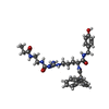  Mass: 615.723 Da / Num. of mol.: 1 / Source method: obtained synthetically / Formula: C33H41N7O5 Mass: 615.723 Da / Num. of mol.: 1 / Source method: obtained synthetically / Formula: C33H41N7O5 |
| Sequence details | Authors state that the site (359-366) is the prescission protease site connected to the C terminal ...Authors state that the site (359-366) is the prescission protease site connected to the C terminal of the receptor. |
-Experimental details
-Experiment
| Experiment | Method: X-RAY DIFFRACTION / Number of used crystals: 1 |
|---|
- Sample preparation
| Crystal | Density Matthews: 2.58 Å3/Da / Density % sol: 52.34 % |
|---|---|
| Crystal grow | Temperature: 293 K / Method: lipidic cubic phase / pH: 7.4 Details: 0.1 M Tris, pH 7.4-8.0, 30-40% (v/v) PEG400, 50-150 mM sodium tartrate and 100 uM UR-MK299 PH range: 7.4-8.0 |
-Data collection
| Diffraction | Mean temperature: 100 K |
|---|---|
| Diffraction source | Source: SYNCHROTRON / Site: SPring-8  / Beamline: BL41XU / Wavelength: 1 Å / Beamline: BL41XU / Wavelength: 1 Å |
| Detector | Type: DECTRIS PILATUS3 6M / Detector: PIXEL / Date: Jan 21, 2016 |
| Radiation | Protocol: SINGLE WAVELENGTH / Monochromatic (M) / Laue (L): M / Scattering type: x-ray |
| Radiation wavelength | Wavelength: 1 Å / Relative weight: 1 |
| Reflection | Resolution: 2.7→50 Å / Num. obs: 16689 / % possible obs: 97.3 % / Redundancy: 3.97 % / Biso Wilson estimate: 78.39 Å2 / CC1/2: 0.988 / Rmerge(I) obs: 0.166 / Net I/σ(I): 4.78 |
| Reflection shell | Resolution: 2.7→2.83 Å / Redundancy: 3.64 % / Rmerge(I) obs: 0.937 / Mean I/σ(I) obs: 1 / Num. unique obs: 2119 / CC1/2: 0.633 / % possible all: 96.9 |
- Processing
| Software |
| ||||||||||||||||||||||||||||||||||||||||||||||||||||||||||||||||||||||||||||||||||||||||||||||||||||||||||||||||||
|---|---|---|---|---|---|---|---|---|---|---|---|---|---|---|---|---|---|---|---|---|---|---|---|---|---|---|---|---|---|---|---|---|---|---|---|---|---|---|---|---|---|---|---|---|---|---|---|---|---|---|---|---|---|---|---|---|---|---|---|---|---|---|---|---|---|---|---|---|---|---|---|---|---|---|---|---|---|---|---|---|---|---|---|---|---|---|---|---|---|---|---|---|---|---|---|---|---|---|---|---|---|---|---|---|---|---|---|---|---|---|---|---|---|---|---|
| Refinement | Method to determine structure: MOLECULAR REPLACEMENT Starting model: 4GRV, 1C5P Resolution: 2.7→29.38 Å / Cor.coef. Fo:Fc: 0.903 / Cor.coef. Fo:Fc free: 0.876 / Rfactor Rfree error: 0 / SU R Cruickshank DPI: 0.974 / Cross valid method: THROUGHOUT / σ(F): 0 / SU R Blow DPI: 0.745 / SU Rfree Blow DPI: 0.306 / SU Rfree Cruickshank DPI: 0.32
| ||||||||||||||||||||||||||||||||||||||||||||||||||||||||||||||||||||||||||||||||||||||||||||||||||||||||||||||||||
| Displacement parameters | Biso mean: 87.93 Å2
| ||||||||||||||||||||||||||||||||||||||||||||||||||||||||||||||||||||||||||||||||||||||||||||||||||||||||||||||||||
| Refine analyze | Luzzati coordinate error obs: 0.47 Å | ||||||||||||||||||||||||||||||||||||||||||||||||||||||||||||||||||||||||||||||||||||||||||||||||||||||||||||||||||
| Refinement step | Cycle: 1 / Resolution: 2.7→29.38 Å
| ||||||||||||||||||||||||||||||||||||||||||||||||||||||||||||||||||||||||||||||||||||||||||||||||||||||||||||||||||
| Refine LS restraints |
| ||||||||||||||||||||||||||||||||||||||||||||||||||||||||||||||||||||||||||||||||||||||||||||||||||||||||||||||||||
| LS refinement shell | Resolution: 2.7→2.89 Å / Rfactor Rfree error: 0 / Total num. of bins used: 8
| ||||||||||||||||||||||||||||||||||||||||||||||||||||||||||||||||||||||||||||||||||||||||||||||||||||||||||||||||||
| Refinement TLS params. | Method: refined / Origin x: -50.2552 Å / Origin y: -14.9395 Å / Origin z: 95.5975 Å
| ||||||||||||||||||||||||||||||||||||||||||||||||||||||||||||||||||||||||||||||||||||||||||||||||||||||||||||||||||
| Refinement TLS group | Selection details: { A|* } |
