 Movie
Movie Controller

[English] 日本語
 Yorodumi
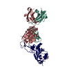
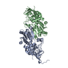
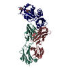
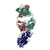
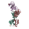
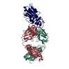
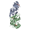
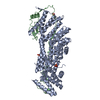
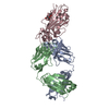
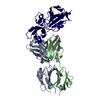
- PDB-7b3o: Crystal structure of the SARS-CoV-2 RBD in complex with STE90-C11 Fab -
+ Open data

- Basic information
| Entry | Database: PDB / ID: 7b3o | ||||||
|---|---|---|---|---|---|---|---|
| Title | Crystal structure of the SARS-CoV-2 RBD in complex with STE90-C11 Fab | ||||||
 Components Components |
| ||||||
 Keywords Keywords | ANTIVIRAL PROTEIN / neutralizing antibodies / SARS-CoV-2 / RBD | ||||||
| Function / homology |  Function and homology information Function and homology informationsymbiont-mediated disruption of host tissue / Maturation of spike protein / Translation of Structural Proteins / Virion Assembly and Release / host cell surface / host extracellular region / symbiont-mediated-mediated suppression of host tetherin activity / Induction of Cell-Cell Fusion / structural constituent of virion / positive regulation of viral entry into host cell ...symbiont-mediated disruption of host tissue / Maturation of spike protein / Translation of Structural Proteins / Virion Assembly and Release / host cell surface / host extracellular region / symbiont-mediated-mediated suppression of host tetherin activity / Induction of Cell-Cell Fusion / structural constituent of virion / positive regulation of viral entry into host cell / membrane fusion / host cell endoplasmic reticulum-Golgi intermediate compartment membrane / Attachment and Entry / entry receptor-mediated virion attachment to host cell / receptor-mediated virion attachment to host cell / host cell surface receptor binding / symbiont-mediated suppression of host innate immune response / endocytosis involved in viral entry into host cell / receptor ligand activity / fusion of virus membrane with host plasma membrane / fusion of virus membrane with host endosome membrane / viral envelope / symbiont entry into host cell / virion attachment to host cell / host cell plasma membrane / SARS-CoV-2 activates/modulates innate and adaptive immune responses / virion membrane / membrane / identical protein binding / plasma membrane Similarity search - Function | ||||||
| Biological species |   Severe acute respiratory syndrome coronavirus 2 Severe acute respiratory syndrome coronavirus 2 Homo sapiens (human) Homo sapiens (human) | ||||||
| Method |  X-RAY DIFFRACTION / SYNCHROTRON / MOLECULAR REPLACEMENT / molecular replacement / Resolution: 2 Å X-RAY DIFFRACTION / SYNCHROTRON / MOLECULAR REPLACEMENT / molecular replacement / Resolution: 2 Å | ||||||
 Authors Authors | Kluenemann, T. / Van den Heuvel, J. | ||||||
| Funding support |  Germany, 1items Germany, 1items
| ||||||
 Citation Citation | Journal: Cell Rep / Year: 2021 Title: A SARS-CoV-2 neutralizing antibody selected from COVID-19 patients binds to the ACE2-RBD interface and is tolerant to most known RBD mutations. Authors: Bertoglio, F. / Fuhner, V. / Ruschig, M. / Heine, P.A. / Abassi, L. / Klunemann, T. / Rand, U. / Meier, D. / Langreder, N. / Steinke, S. / Ballmann, R. / Schneider, K.T. / Roth, K.D.R. / ...Authors: Bertoglio, F. / Fuhner, V. / Ruschig, M. / Heine, P.A. / Abassi, L. / Klunemann, T. / Rand, U. / Meier, D. / Langreder, N. / Steinke, S. / Ballmann, R. / Schneider, K.T. / Roth, K.D.R. / Kuhn, P. / Riese, P. / Schackermann, D. / Korn, J. / Koch, A. / Chaudhry, M.Z. / Eschke, K. / Kim, Y. / Zock-Emmenthal, S. / Becker, M. / Scholz, M. / Moreira, G.M.S.G. / Wenzel, E.V. / Russo, G. / Garritsen, H.S.P. / Casu, S. / Gerstner, A. / Roth, G. / Adler, J. / Trimpert, J. / Hermann, A. / Schirrmann, T. / Dubel, S. / Frenzel, A. / Van den Heuvel, J. / Cicin-Sain, L. / Schubert, M. / Hust, M. #1: Journal: Cell Rep / Year: 2021Title: A SARS-CoV-2 neutralizing antibody selected from COVID-19 patients binds to the ACE2-RBD interface and is tolerant to most known RBD mutations. Authors: Bertoglio, F. / Fuhner, V. / Ruschig, M. / Heine, P.A. / Abassi, L. / Klunemann, T. / Rand, U. / Meier, D. / Langreder, N. / Steinke, S. / Ballmann, R. / Schneider, K.T. / Roth, K.D.R. / ...Authors: Bertoglio, F. / Fuhner, V. / Ruschig, M. / Heine, P.A. / Abassi, L. / Klunemann, T. / Rand, U. / Meier, D. / Langreder, N. / Steinke, S. / Ballmann, R. / Schneider, K.T. / Roth, K.D.R. / Kuhn, P. / Riese, P. / Schackermann, D. / Korn, J. / Koch, A. / Chaudhry, M.Z. / Eschke, K. / Kim, Y. / Zock-Emmenthal, S. / Becker, M. / Scholz, M. / Moreira, G.M.S.G. / Wenzel, E.V. / Russo, G. / Garritsen, H.S.P. / Casu, S. / Gerstner, A. / Roth, G. / Adler, J. / Trimpert, J. / Hermann, A. / Schirrmann, T. / Dubel, S. / Frenzel, A. / Van den Heuvel, J. / Cicin-Sain, L. / Schubert, M. / Hust, M. #2: Journal: Biorxiv / Year: 2020Title: A SARS-CoV-2 neutralizing antibody selected from COVID-19 patients by phage display is binding to the ACE2-RBD interface and is tolerant to known RBD mutations Authors: Bertoglio, F. / Fuhner, V. / Ruschig, M. / Heine, P.A. / Rand, U. / Klunemann, T. / Meier, D. / Langreder, N. / Steinke, S. / Ballmann, R. / Schneider, K. / Roth, K.D.R. / Kuhn, P. / Riese, ...Authors: Bertoglio, F. / Fuhner, V. / Ruschig, M. / Heine, P.A. / Rand, U. / Klunemann, T. / Meier, D. / Langreder, N. / Steinke, S. / Ballmann, R. / Schneider, K. / Roth, K.D.R. / Kuhn, P. / Riese, P. / Schackermann, D. / Korn, J. / Koch, A. / Zock-Emmenthal, S. / Becker, M. / Scholz, M. / Moreira, G.M.S.G. / Wenzel, E.V. / Russo, G. / Garritsen, H.S. / Casu, S. / Gerstner, A. / Roth, G. / Hermann, A. / Schirrmann, T. / Dubel, S. / Frenzel, A. / Cicin-Sain, L. / Schubert, M. / Hust, M. | ||||||
| History |
|
- Structure visualization
| Structure viewer | Molecule: MolmilJmol/JSmol |
|---|
- Downloads & links
-Download
| PDBx/mmCIF format | 7b3o.cif.gz | 243.9 KB | Display | PDBx/mmCIF format |
|---|---|---|---|---|
| PDB format | pdb7b3o.ent.gz | 195.1 KB | Display | PDB format |
| PDBx/mmJSON format | 7b3o.json.gz | Tree view | PDBx/mmJSON format | |
| Others |  Other downloads Other downloads |
-Validation report
| Arichive directory | https://data.pdbj.org/pub/pdb/validation_reports/b3/7b3oftp://data.pdbj.org/pub/pdb/validation_reports/b3/7b3o | HTTPS FTP |
|---|
-Related structure data
| Related structure data | 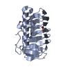 7bwyS S: Starting model for refinement |
|---|---|
| Similar structure data |
-Links
 PDBj
PDBj


- Assembly
| Deposited unit | 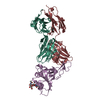
| ||||||||
|---|---|---|---|---|---|---|---|---|---|
| 1 |
| ||||||||
| Unit cell |
| ||||||||
| Components on special symmetry positions |
|
-Components
| #1: Protein | Mass: 23087.838 Da / Num. of mol.: 1 Source method: isolated from a genetically manipulated source Source: (gene. exp.) Severe acute respiratory syndrome coronavirus 2Gene: S, 2 / Production host:  Trichoplusia ni (cabbage looper) / References: UniProt: P0DTC2 Trichoplusia ni (cabbage looper) / References: UniProt: P0DTC2 |
|---|---|
| #2: Antibody | Mass: 23481.197 Da / Num. of mol.: 1 Source method: isolated from a genetically manipulated source Source: (gene. exp.) Homo sapiens (human) / Production host: Homo sapiens (human) |
| #3: Antibody | Mass: 24081.908 Da / Num. of mol.: 1 Source method: isolated from a genetically manipulated source Source: (gene. exp.) Homo sapiens (human) / Production host: Homo sapiens (human) |
| #4: Polysaccharide | 2-acetamido-2-deoxy-beta-D-glucopyranose-(1-4)-2-acetamido-2-deoxy-beta-D-glucopyranose Source method: isolated from a genetically manipulated source |
| #5: Water | ChemComp-HOH /  Mass: 18.015 Da / Num. of mol.: 427 / Source method: isolated from a natural source / Formula: H2O Mass: 18.015 Da / Num. of mol.: 427 / Source method: isolated from a natural source / Formula: H2O |
| Has ligand of interest | N |
| Has protein modification | Y |
-Experimental details
-Experiment
| Experiment | Method: X-RAY DIFFRACTION / Number of used crystals: 1 |
|---|
- Sample preparation
| Crystal | Density Matthews: 3.4 Å3/Da / Density % sol: 63.78 % |
|---|---|
| Crystal grow | Temperature: 298 K / Method: vapor diffusion Details: 13.3% (w/v) polyethylene glycol 6,000, 0.1M MES pH 5.6 0.24M tri sodium citrate |
-Data collection
| Diffraction | Mean temperature: 100 K / Serial crystal experiment: N | ||||||||||||||||||||||||||||||
|---|---|---|---|---|---|---|---|---|---|---|---|---|---|---|---|---|---|---|---|---|---|---|---|---|---|---|---|---|---|---|---|
| Diffraction source | Source: SYNCHROTRON / Site: PETRA III, DESY / Beamline: P11 / Wavelength: 1.033 Å | ||||||||||||||||||||||||||||||
| Detector | Type: DECTRIS PILATUS 6M-F / Detector: PIXEL / Date: Sep 4, 2020 | ||||||||||||||||||||||||||||||
| Radiation | Protocol: SINGLE WAVELENGTH / Monochromatic (M) / Laue (L): M / Scattering type: x-ray | ||||||||||||||||||||||||||||||
| Radiation wavelength | Wavelength: 1.033 Å / Relative weight: 1 | ||||||||||||||||||||||||||||||
| Reflection | Resolution: 2→48.1 Å / Num. obs: 63906 / % possible obs: 100 % / Redundancy: 6.9 % / CC1/2: 0.985 / Rmerge(I) obs: 0.159 / Rpim(I) all: 0.066 / Rrim(I) all: 0.173 / Net I/σ(I): 6.7 / Num. measured all: 437879 / Scaling rejects: 33 | ||||||||||||||||||||||||||||||
| Reflection shell | Diffraction-ID: 1
|
-Phasing
| Phasing | Method: molecular replacement | |||||||||
|---|---|---|---|---|---|---|---|---|---|---|
| Phasing MR |
|
- Processing
| Software |
| ||||||||||||||||||||||||||||||||||||||||||||||||||||||
|---|---|---|---|---|---|---|---|---|---|---|---|---|---|---|---|---|---|---|---|---|---|---|---|---|---|---|---|---|---|---|---|---|---|---|---|---|---|---|---|---|---|---|---|---|---|---|---|---|---|---|---|---|---|---|---|
| Refinement | Method to determine structure: MOLECULAR REPLACEMENT Starting model: 7bwy Resolution: 2→48.1 Å / SU ML: 0.28 / Cross valid method: THROUGHOUT / σ(F): 1.34 / Phase error: 26.87 / Stereochemistry target values: ML
| ||||||||||||||||||||||||||||||||||||||||||||||||||||||
| Solvent computation | Shrinkage radii: 0.9 Å / VDW probe radii: 1.11 Å / Solvent model: FLAT BULK SOLVENT MODEL | ||||||||||||||||||||||||||||||||||||||||||||||||||||||
| Displacement parameters | Biso max: 160.82 Å2 / Biso mean: 47.2921 Å2 / Biso min: 22.59 Å2 | ||||||||||||||||||||||||||||||||||||||||||||||||||||||
| Refinement step | Cycle: final / Resolution: 2→48.1 Å
| ||||||||||||||||||||||||||||||||||||||||||||||||||||||
| LS refinement shell | Refine-ID: X-RAY DIFFRACTION / Rfactor Rfree error: 0 / Total num. of bins used: 8 / % reflection obs: 100 %
|
