 Movie
Movie Controller

[English] 日本語
 Yorodumi
- PDB-5z7j: Crystal structure of a lactonase double mutant in complex with li... -
+ Open data

- Basic information
| Entry | Database: PDB / ID: 5z7j | ||||||
|---|---|---|---|---|---|---|---|
| Title | Crystal structure of a lactonase double mutant in complex with ligand l | ||||||
 Components Components | Lactonase for protein | ||||||
 Keywords Keywords | HYDROLASE / ALPHA/BETA-HYDROLASE / LACTONASE / ZEARALENONE | ||||||
| Function / homology | : / alpha/beta hydrolase fold / Alpha/beta hydrolase fold-1 / Alpha/Beta hydrolase fold / Chem-36J / DI(HYDROXYETHYL)ETHER / AB hydrolase-1 domain-containing protein Function and homology information Function and homology information | ||||||
| Biological species |  Rhinocladiella mackenziei CBS 650.93 (fungus) Rhinocladiella mackenziei CBS 650.93 (fungus) | ||||||
| Method |  X-RAY DIFFRACTION / SYNCHROTRON / MOLECULAR REPLACEMENT / Resolution: 1.98 Å X-RAY DIFFRACTION / SYNCHROTRON / MOLECULAR REPLACEMENT / Resolution: 1.98 Å | ||||||
 Authors Authors | Zheng, Y.Y. / Liu, W.D. / Chen, C.C. / Guo, R.T. | ||||||
| Funding support |  China, 1items China, 1items
| ||||||
 Citation Citation | Journal: Acs Catalysis / Year: 2018 Title: Crystal Structure of a Mycoestrogen-Detoxifying Lactonase from Rhinocladiella mackenziei: Molecular Insight into ZHD Substrate Selectivity Authors: Zheng, Y.Y. / Liu, W.T. / Chen, C.C. / Hu, X.Y. / Liu, W.D. / Ko, T.P. / Tang, X.K. / Wei, H.L. / Huang, J.W. / Guo, R.T. | ||||||
| History |
|
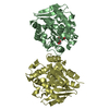
- Structure visualization
| Structure viewer | Molecule: MolmilJmol/JSmol |
|---|
- Downloads & links
-Download
| PDBx/mmCIF format | 5z7j.cif.gz | 450.6 KB | Display | PDBx/mmCIF format |
|---|---|---|---|---|
| PDB format | pdb5z7j.ent.gz | 368.1 KB | Display | PDB format |
| PDBx/mmJSON format | 5z7j.json.gz | Tree view | PDBx/mmJSON format | |
| Others |  Other downloads Other downloads |
-Validation report
| Arichive directory | https://data.pdbj.org/pub/pdb/validation_reports/z7/5z7jftp://data.pdbj.org/pub/pdb/validation_reports/z7/5z7j | HTTPS FTP |
|---|
-Related structure data
| Related structure data | 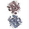 5xo6C 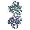 5xo7C 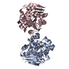 5xo8C  5z5jC  5z97C  5ie4S S: Starting model for refinement C: citing same article ( |
|---|---|
| Similar structure data |


-Links
 PDBj
PDBj


- Assembly
| Deposited unit | 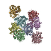
| ||||||||
|---|---|---|---|---|---|---|---|---|---|
| 1 | 
| ||||||||
| 2 | 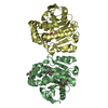
| ||||||||
| 3 | 
| ||||||||
| 4 | 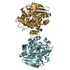
| ||||||||
| Unit cell |
|
-Components
| #1: Protein | Mass: 29154.854 Da / Num. of mol.: 8 / Fragment: UNP residues 3-266 Source method: isolated from a genetically manipulated source Source: (gene. exp.) Rhinocladiella mackenziei CBS 650.93 (fungus)Gene: Z518_04590 / Plasmid: pET-46 EK/LIC / Production host:  #2: Chemical | ChemComp-36J / ( 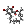  Mass: 320.380 Da / Num. of mol.: 8 / Source method: obtained synthetically / Formula: C18H24O5 Mass: 320.380 Da / Num. of mol.: 8 / Source method: obtained synthetically / Formula: C18H24O5#3: Chemical |   Mass: 106.120 Da / Num. of mol.: 2 / Source method: obtained synthetically / Formula: C4H10O3 Mass: 106.120 Da / Num. of mol.: 2 / Source method: obtained synthetically / Formula: C4H10O3#4: Water | ChemComp-HOH / |  Mass: 18.015 Da / Num. of mol.: 1864 / Source method: isolated from a natural source / Formula: H2O Mass: 18.015 Da / Num. of mol.: 1864 / Source method: isolated from a natural source / Formula: H2O |
|---|
-Experimental details
-Experiment
| Experiment | Method: X-RAY DIFFRACTION / Number of used crystals: 1 |
|---|
- Sample preparation
| Crystal | Density Matthews: 3.07 Å3/Da / Density % sol: 59.94 % / Mosaicity: 0.908 ° |
|---|---|
| Crystal grow | Temperature: 298 K / Method: vapor diffusion, sitting drop / pH: 6.5 Details: 0.2M Ammonium Sulfate, 0.085M Sodium Cacodylate pH 6.5, 25-28%(w/v) Polyethylene Glycol 8000 and 15%(v/v) Glycerol |
-Data collection
| Diffraction | Mean temperature: 100 K | |||||||||||||||||||||||||||||||||||||||||||||||||||||||||||||||||||||||||||||||||||||||||||||||||||
|---|---|---|---|---|---|---|---|---|---|---|---|---|---|---|---|---|---|---|---|---|---|---|---|---|---|---|---|---|---|---|---|---|---|---|---|---|---|---|---|---|---|---|---|---|---|---|---|---|---|---|---|---|---|---|---|---|---|---|---|---|---|---|---|---|---|---|---|---|---|---|---|---|---|---|---|---|---|---|---|---|---|---|---|---|---|---|---|---|---|---|---|---|---|---|---|---|---|---|---|---|
| Diffraction source | Source: SYNCHROTRON / Site: NSRRC  / Beamline: BL15A1 / Wavelength: 1 Å / Beamline: BL15A1 / Wavelength: 1 Å | |||||||||||||||||||||||||||||||||||||||||||||||||||||||||||||||||||||||||||||||||||||||||||||||||||
| Detector | Type: RAYONIX MX-300 / Detector: CCD / Date: Aug 2, 2017 | |||||||||||||||||||||||||||||||||||||||||||||||||||||||||||||||||||||||||||||||||||||||||||||||||||
| Radiation | Monochromator: GRAPHITE / Protocol: SINGLE WAVELENGTH / Monochromatic (M) / Laue (L): M / Scattering type: x-ray | |||||||||||||||||||||||||||||||||||||||||||||||||||||||||||||||||||||||||||||||||||||||||||||||||||
| Radiation wavelength | Wavelength: 1 Å / Relative weight: 1 | |||||||||||||||||||||||||||||||||||||||||||||||||||||||||||||||||||||||||||||||||||||||||||||||||||
| Reflection | Resolution: 1.98→25 Å / Num. obs: 188099 / % possible obs: 97.7 % / Redundancy: 4 % / Biso Wilson estimate: 30.94 Å2 / Rmerge(I) obs: 0.073 / Rpim(I) all: 0.042 / Rrim(I) all: 0.085 / Χ2: 0.705 / Net I/σ(I): 10.6 / Num. measured all: 752400 | |||||||||||||||||||||||||||||||||||||||||||||||||||||||||||||||||||||||||||||||||||||||||||||||||||
| Reflection shell | Diffraction-ID: 1
|
- Processing
| Software |
| |||||||||||||||||||||||||||||||||||||||||||||||||||||||||||||||||||||||||||||
|---|---|---|---|---|---|---|---|---|---|---|---|---|---|---|---|---|---|---|---|---|---|---|---|---|---|---|---|---|---|---|---|---|---|---|---|---|---|---|---|---|---|---|---|---|---|---|---|---|---|---|---|---|---|---|---|---|---|---|---|---|---|---|---|---|---|---|---|---|---|---|---|---|---|---|---|---|---|---|
| Refinement | Method to determine structure: MOLECULAR REPLACEMENT Starting model: 5IE4 Resolution: 1.98→24.714 Å / SU ML: 0.22 / Cross valid method: THROUGHOUT / σ(F): 1.96 / Phase error: 22.46
| |||||||||||||||||||||||||||||||||||||||||||||||||||||||||||||||||||||||||||||
| Solvent computation | Shrinkage radii: 0.9 Å / VDW probe radii: 1.11 Å | |||||||||||||||||||||||||||||||||||||||||||||||||||||||||||||||||||||||||||||
| Displacement parameters | Biso max: 90.35 Å2 / Biso mean: 36.0956 Å2 / Biso min: 18.93 Å2 | |||||||||||||||||||||||||||||||||||||||||||||||||||||||||||||||||||||||||||||
| Refinement step | Cycle: final / Resolution: 1.98→24.714 Å
| |||||||||||||||||||||||||||||||||||||||||||||||||||||||||||||||||||||||||||||
| Refine LS restraints |
| |||||||||||||||||||||||||||||||||||||||||||||||||||||||||||||||||||||||||||||
| LS refinement shell | Refine-ID: X-RAY DIFFRACTION / Rfactor Rfree error: 0 / Total num. of bins used: 10
|
