 Movie
Movie Controller


+ Open data

- Basic information
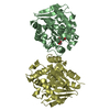
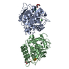
| Entry | Database: PDB / ID: 5xo7 | ||||||
|---|---|---|---|---|---|---|---|
| Title | Crystal structure of a novel ZEN lactonase mutant with ligand a | ||||||
 Components Components | lactonase for protein | ||||||
 Keywords Keywords | HYDROLASE / ALPHA/BETA-HYDROLASE / LACTONASE / ZEARALENONE | ||||||
| Function / homology | : / alpha/beta hydrolase fold / Alpha/beta hydrolase fold-1 / Alpha/Beta hydrolase fold / Chem-36J / AB hydrolase-1 domain-containing protein Function and homology information Function and homology information | ||||||
| Biological species |  Rhinocladiella mackenziei CBS 650.93 (fungus) Rhinocladiella mackenziei CBS 650.93 (fungus) | ||||||
| Method |  X-RAY DIFFRACTION / SYNCHROTRON / MOLECULAR REPLACEMENT / Resolution: 1.88 Å X-RAY DIFFRACTION / SYNCHROTRON / MOLECULAR REPLACEMENT / Resolution: 1.88 Å | ||||||
 Authors Authors | Zheng, Y.Y. / Liu, W.T. / Liu, W.D. / Chen, C.C. / Guo, R.T. | ||||||
 Citation Citation | Journal: Acs Catalysis / Year: 2018 Title: Crystal Structure of a Mycoestrogen-Detoxifying Lactonase from Rhinocladiella mackenziei: Molecular Insight into ZHD Substrate Selectivity Authors: Zheng, Y.Y. / Liu, W.T. / Chen, C.C. / Hu, X.Y. / Liu, W.D. / Ko, T.P. / Tang, X.K. / Wei, H.L. / Huang, J.W. / Guo, R.T. | ||||||
| History |
|
- Structure visualization
| Structure viewer | Molecule: MolmilJmol/JSmol |
|---|
- Downloads & links
-Download
| PDBx/mmCIF format | 5xo7.cif.gz | 455.7 KB | Display | PDBx/mmCIF format |
|---|---|---|---|---|
| PDB format | pdb5xo7.ent.gz | 376.3 KB | Display | PDB format |
| PDBx/mmJSON format | 5xo7.json.gz | Tree view | PDBx/mmJSON format | |
| Others |  Other downloads Other downloads |
-Validation report
| Arichive directory | https://data.pdbj.org/pub/pdb/validation_reports/xo/5xo7ftp://data.pdbj.org/pub/pdb/validation_reports/xo/5xo7 | HTTPS FTP |
|---|
-Related structure data
| Related structure data |  5xo6C  5xo8C  5z5jC  5z7jC  5z97C  3wzlS C: citing same article ( S: Starting model for refinement |
|---|---|
| Similar structure data |


-Links
 PDBj
PDBj


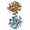
- Assembly
| Deposited unit | 
| ||||||||
|---|---|---|---|---|---|---|---|---|---|
| 1 | 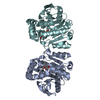
| ||||||||
| 2 | 
| ||||||||
| 3 | 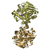
| ||||||||
| 4 | 
| ||||||||
| Unit cell |
|
-Components
| #1: Protein | Mass: 29449.219 Da / Num. of mol.: 8 / Mutation: S105A Source method: isolated from a genetically manipulated source Source: (gene. exp.) Rhinocladiella mackenziei CBS 650.93 (fungus)Gene: Z518_04590 / Plasmid: pET-46 EK/LIC / Production host:  #2: Chemical | ChemComp-36J / ( 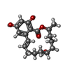  Mass: 320.380 Da / Num. of mol.: 8 / Source method: obtained synthetically / Formula: C18H24O5 Mass: 320.380 Da / Num. of mol.: 8 / Source method: obtained synthetically / Formula: C18H24O5#3: Water | ChemComp-HOH / |  Mass: 18.015 Da / Num. of mol.: 2210 / Source method: isolated from a natural source / Formula: H2O Mass: 18.015 Da / Num. of mol.: 2210 / Source method: isolated from a natural source / Formula: H2O |
|---|
-Experimental details
-Experiment
| Experiment | Method: X-RAY DIFFRACTION / Number of used crystals: 1 |
|---|
- Sample preparation
| Crystal | Density Matthews: 3.09 Å3/Da / Density % sol: 60.15 % / Mosaicity: 0.277 ° |
|---|---|
| Crystal grow | Temperature: 298 K / Method: vapor diffusion, sitting drop / pH: 6.5 Details: 0.2M Ammonium Sulfate, 0.085M Sodium Cacodylate pH 6.5, 25-28%(w/v) Polyethylene Glycol 8000 and 15%(v/v) Glycerol |
-Data collection
| Diffraction | Mean temperature: 100 K |
|---|---|
| Diffraction source | Source: SYNCHROTRON / Site: NSRRC  / Beamline: TPS 05A / Wavelength: 0.9998 Å / Beamline: TPS 05A / Wavelength: 0.9998 Å |
| Detector | Type: RAYONIX MX300-HS / Detector: CCD / Date: Apr 16, 2017 |
| Radiation | Monochromator: LN2 cooled Si(111) double crystal monochromator Protocol: SINGLE WAVELENGTH / Monochromatic (M) / Laue (L): M / Scattering type: x-ray |
| Radiation wavelength | Wavelength: 0.9998 Å / Relative weight: 1 |
| Reflection | Resolution: 1.88→25 Å / Num. obs: 222958 / % possible obs: 97.6 % / Redundancy: 3.9 % / Rmerge(I) obs: 0.08 / Net I/σ(I): 6.8 |
| Reflection shell | Resolution: 1.88→1.95 Å / Redundancy: 3.7 % / Rmerge(I) obs: 0.498 / % possible all: 94.9 |
- Processing
| Software |
| ||||||||||||||||||||||||||||||||||||||||||||||||||||||||||||||||||||||||||||||||||||||||||||||||||||||||||||||||||||||||||||||||||||||||||||||||||||||||||||||||||||||||||||||||||||||
|---|---|---|---|---|---|---|---|---|---|---|---|---|---|---|---|---|---|---|---|---|---|---|---|---|---|---|---|---|---|---|---|---|---|---|---|---|---|---|---|---|---|---|---|---|---|---|---|---|---|---|---|---|---|---|---|---|---|---|---|---|---|---|---|---|---|---|---|---|---|---|---|---|---|---|---|---|---|---|---|---|---|---|---|---|---|---|---|---|---|---|---|---|---|---|---|---|---|---|---|---|---|---|---|---|---|---|---|---|---|---|---|---|---|---|---|---|---|---|---|---|---|---|---|---|---|---|---|---|---|---|---|---|---|---|---|---|---|---|---|---|---|---|---|---|---|---|---|---|---|---|---|---|---|---|---|---|---|---|---|---|---|---|---|---|---|---|---|---|---|---|---|---|---|---|---|---|---|---|---|---|---|---|---|
| Refinement | Method to determine structure: MOLECULAR REPLACEMENT Starting model: 3WZL Resolution: 1.88→24.93 Å / Cor.coef. Fo:Fc: 0.971 / Cor.coef. Fo:Fc free: 0.956 / SU B: 3.046 / SU ML: 0.087 / Cross valid method: THROUGHOUT / ESU R: 0.107 / ESU R Free: 0.109 / Stereochemistry target values: MAXIMUM LIKELIHOOD / Details: HYDROGENS HAVE BEEN ADDED IN THE RIDING POSITIONS
| ||||||||||||||||||||||||||||||||||||||||||||||||||||||||||||||||||||||||||||||||||||||||||||||||||||||||||||||||||||||||||||||||||||||||||||||||||||||||||||||||||||||||||||||||||||||
| Solvent computation | Ion probe radii: 0.8 Å / Shrinkage radii: 0.8 Å / VDW probe radii: 1.2 Å / Solvent model: MASK | ||||||||||||||||||||||||||||||||||||||||||||||||||||||||||||||||||||||||||||||||||||||||||||||||||||||||||||||||||||||||||||||||||||||||||||||||||||||||||||||||||||||||||||||||||||||
| Displacement parameters | Biso mean: 31.24 Å2
| ||||||||||||||||||||||||||||||||||||||||||||||||||||||||||||||||||||||||||||||||||||||||||||||||||||||||||||||||||||||||||||||||||||||||||||||||||||||||||||||||||||||||||||||||||||||
| Refinement step | Cycle: LAST / Resolution: 1.88→24.93 Å
| ||||||||||||||||||||||||||||||||||||||||||||||||||||||||||||||||||||||||||||||||||||||||||||||||||||||||||||||||||||||||||||||||||||||||||||||||||||||||||||||||||||||||||||||||||||||
| Refine LS restraints |
|