 Movie
Movie Controller

[English] 日本語
 Yorodumi
- PDB-5ie4: Crystal structure of a lactonase mutant in complex with substrate a -
+ Open data

- Basic information
| Entry | Database: PDB / ID: 5ie4 | ||||||
|---|---|---|---|---|---|---|---|
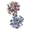
| Title | Crystal structure of a lactonase mutant in complex with substrate a | ||||||
 Components Components | Zearalenone hydrolase | ||||||
 Keywords Keywords | HYDROLASE / ALPHA/BETA-HYDROLASE / LACTONASE / ZEARALENONE | ||||||
| Function / homology | : / Hydrolases; Acting on ester bonds; Carboxylic-ester hydrolases / alpha/beta hydrolase fold / Alpha/beta hydrolase fold-1 / response to toxic substance / Alpha/Beta hydrolase fold / hydrolase activity / Chem-36J / Zearalenone hydrolase Function and homology information Function and homology information | ||||||
| Biological species |  Clonostachys rosea (fungus) Clonostachys rosea (fungus) | ||||||
| Method |  X-RAY DIFFRACTION / SYNCHROTRON / MOLECULAR REPLACEMENT / molecular replacement / Resolution: 2.8 Å X-RAY DIFFRACTION / SYNCHROTRON / MOLECULAR REPLACEMENT / molecular replacement / Resolution: 2.8 Å | ||||||
 Authors Authors | Zheng, Y.Y. / Xu, Z.X. / Liu, W.D. / Chen, C.C. / Guo, R.T. | ||||||
 Citation Citation | Journal: Acs Catalysis / Year: 2016 Title: Enhanced alph-Zearalenol Hydrolyzing Activity of a Mycoestrogen-Detoxifying Lactonase by Structure-Based Engineering Authors: Xu, Z.X. / Liu, W.D. / Chen, C.C. / Li, Q. / Huang, J.W. / Ko, T.P. / Liu, G. / Liu, W. / Peng, W. / Cheng, Y.S. / Chen, Y. / Jin, J. / Li, H. / Zheng, Y.Y. / Guo, R.T. | ||||||
| History |
|
- Structure visualization
| Structure viewer | Molecule: MolmilJmol/JSmol |
|---|
- Downloads & links
-Download
| PDBx/mmCIF format | 5ie4.cif.gz | 147.5 KB | Display | PDBx/mmCIF format |
|---|---|---|---|---|
| PDB format | pdb5ie4.ent.gz | 115.2 KB | Display | PDB format |
| PDBx/mmJSON format | 5ie4.json.gz | Tree view | PDBx/mmJSON format | |
| Others |  Other downloads Other downloads |
-Validation report
| Arichive directory | https://data.pdbj.org/pub/pdb/validation_reports/ie/5ie4ftp://data.pdbj.org/pub/pdb/validation_reports/ie/5ie4 | HTTPS FTP |
|---|
-Related structure data
| Related structure data |  5ie5C  5ie6C  5ie7C  3wzlS C: citing same article ( S: Starting model for refinement |
|---|---|
| Similar structure data |


-Links
 PDBj
PDBj


- Assembly
| Deposited unit | 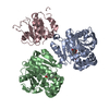
| ||||||||||||||||||
|---|---|---|---|---|---|---|---|---|---|---|---|---|---|---|---|---|---|---|---|
| 1 | 
| ||||||||||||||||||
| 2 | 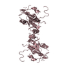
| ||||||||||||||||||
| Unit cell |
| ||||||||||||||||||
| Noncrystallographic symmetry (NCS) | NCS domain:
NCS domain segments: Component-ID: _ / Ens-ID: 1 / Beg auth comp-ID: MET / Beg label comp-ID: MET / End auth comp-ID: LEU / End label comp-ID: LEU / Refine code: _ / Auth seq-ID: 1 - 264 / Label seq-ID: 1 - 264
|
-Components
| #1: Protein | Mass: 28762.627 Da / Num. of mol.: 3 / Mutation: S102A Source method: isolated from a genetically manipulated source Source: (gene. exp.) Clonostachys rosea (fungus) / Gene: zhd101 / Plasmid: pET-46 EK/LIC / Production host:  #2: Chemical |   Mass: 320.380 Da / Num. of mol.: 2 / Source method: obtained synthetically / Formula: C18H24O5 Mass: 320.380 Da / Num. of mol.: 2 / Source method: obtained synthetically / Formula: C18H24O5#3: Water | ChemComp-HOH / |  Mass: 18.015 Da / Num. of mol.: 200 / Source method: isolated from a natural source / Formula: H2O Mass: 18.015 Da / Num. of mol.: 200 / Source method: isolated from a natural source / Formula: H2O |
|---|
-Experimental details
-Experiment
| Experiment | Method: X-RAY DIFFRACTION / Number of used crystals: 1 |
|---|
- Sample preparation
| Crystal | Density Matthews: 2.94 Å3/Da / Density % sol: 58.18 % / Mosaicity: 0.613 ° |
|---|---|
| Crystal grow | Temperature: 298 K / Method: vapor diffusion, sitting drop / pH: 6.5 / Details: 24% PEG 2000 MME, 0.1M Bis-Tris |
-Data collection
| Diffraction | Mean temperature: 100 K | |||||||||||||||||||||||||||||||||||||||||||||||||||||||
|---|---|---|---|---|---|---|---|---|---|---|---|---|---|---|---|---|---|---|---|---|---|---|---|---|---|---|---|---|---|---|---|---|---|---|---|---|---|---|---|---|---|---|---|---|---|---|---|---|---|---|---|---|---|---|---|---|
| Diffraction source | Source: SYNCHROTRON / Site: NSRRC  / Beamline: BL13B1 / Wavelength: 0.97622 Å / Beamline: BL13B1 / Wavelength: 0.97622 Å | |||||||||||||||||||||||||||||||||||||||||||||||||||||||
| Detector | Type: ADSC QUANTUM 315r / Detector: CCD / Date: Jun 20, 2015 | |||||||||||||||||||||||||||||||||||||||||||||||||||||||
| Radiation | Monochromator: GRAPHITE / Protocol: SINGLE WAVELENGTH / Monochromatic (M) / Laue (L): M / Scattering type: x-ray | |||||||||||||||||||||||||||||||||||||||||||||||||||||||
| Radiation wavelength | Wavelength: 0.97622 Å / Relative weight: 1 | |||||||||||||||||||||||||||||||||||||||||||||||||||||||
| Reflection | Resolution: 2.8→25 Å / Num. obs: 26370 / % possible obs: 97.5 % / Redundancy: 6.2 % / Rmerge(I) obs: 0.053 / Rpim(I) all: 0.024 / Rrim(I) all: 0.059 / Χ2: 1.205 / Net I/av σ(I): 36.758 / Net I/σ(I): 13.9 / Num. measured all: 162671 | |||||||||||||||||||||||||||||||||||||||||||||||||||||||
| Reflection shell |
|
-Phasing
| Phasing | Method: molecular replacement |
|---|
- Processing
| Software |
| ||||||||||||||||||||||||||||||||||||||||||||||||||||||||||||||||||||||||||||||||||||||||||||||||||||||||||||||||||||||||||||||||||||||||||||||||||||||||||||||||||||||||||||||||||||||
|---|---|---|---|---|---|---|---|---|---|---|---|---|---|---|---|---|---|---|---|---|---|---|---|---|---|---|---|---|---|---|---|---|---|---|---|---|---|---|---|---|---|---|---|---|---|---|---|---|---|---|---|---|---|---|---|---|---|---|---|---|---|---|---|---|---|---|---|---|---|---|---|---|---|---|---|---|---|---|---|---|---|---|---|---|---|---|---|---|---|---|---|---|---|---|---|---|---|---|---|---|---|---|---|---|---|---|---|---|---|---|---|---|---|---|---|---|---|---|---|---|---|---|---|---|---|---|---|---|---|---|---|---|---|---|---|---|---|---|---|---|---|---|---|---|---|---|---|---|---|---|---|---|---|---|---|---|---|---|---|---|---|---|---|---|---|---|---|---|---|---|---|---|---|---|---|---|---|---|---|---|---|---|---|
| Refinement | Method to determine structure: MOLECULAR REPLACEMENT Starting model: 3WZL Resolution: 2.8→25 Å / Cor.coef. Fo:Fc: 0.924 / Cor.coef. Fo:Fc free: 0.875 / SU B: 22.212 / SU ML: 0.225 / Cross valid method: THROUGHOUT / ESU R: 0.751 / ESU R Free: 0.354 / Stereochemistry target values: MAXIMUM LIKELIHOOD / Details: HYDROGENS HAVE BEEN ADDED IN THE RIDING POSITIONS
| ||||||||||||||||||||||||||||||||||||||||||||||||||||||||||||||||||||||||||||||||||||||||||||||||||||||||||||||||||||||||||||||||||||||||||||||||||||||||||||||||||||||||||||||||||||||
| Solvent computation | Ion probe radii: 0.8 Å / Shrinkage radii: 0.8 Å / VDW probe radii: 1.2 Å / Solvent model: MASK | ||||||||||||||||||||||||||||||||||||||||||||||||||||||||||||||||||||||||||||||||||||||||||||||||||||||||||||||||||||||||||||||||||||||||||||||||||||||||||||||||||||||||||||||||||||||
| Displacement parameters | Biso mean: 36.008 Å2
| ||||||||||||||||||||||||||||||||||||||||||||||||||||||||||||||||||||||||||||||||||||||||||||||||||||||||||||||||||||||||||||||||||||||||||||||||||||||||||||||||||||||||||||||||||||||
| Refinement step | Cycle: LAST / Resolution: 2.8→25 Å
| ||||||||||||||||||||||||||||||||||||||||||||||||||||||||||||||||||||||||||||||||||||||||||||||||||||||||||||||||||||||||||||||||||||||||||||||||||||||||||||||||||||||||||||||||||||||
| Refine LS restraints |
|