 Movie
Movie Controller

[English] 日本語
 Yorodumi
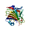
- PDB-2a6x: Crystal structure of Emp46p carbohydrate recognition domain (CRD)... -
+ Open data

- Basic information
| Entry | Database: PDB / ID: 2a6x | ||||||
|---|---|---|---|---|---|---|---|
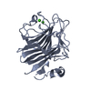
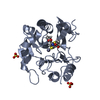
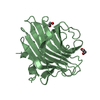
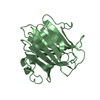
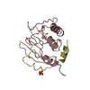
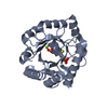
| Title | Crystal structure of Emp46p carbohydrate recognition domain (CRD), Y131F mutant | ||||||
 Components Components | Emp46p | ||||||
 Keywords Keywords | SUGAR BINDING PROTEIN / BETA SANDWICH / CARBOHYDRATE BINDING PROTEIN / CARGO RECEPTOR / Structural Genomics / NPPSFA / National Project on Protein Structural and Functional Analyses / RIKEN Structural Genomics/Proteomics Initiative / RSGI | ||||||
| Function / homology |  Function and homology information Function and homology informationCOPII-coated ER to Golgi transport vesicle / D-mannose binding / endoplasmic reticulum-Golgi intermediate compartment / endoplasmic reticulum to Golgi vesicle-mediated transport / protein transport / carbohydrate binding / Golgi membrane / endoplasmic reticulum membrane / endoplasmic reticulum / metal ion binding Similarity search - Function | ||||||
| Biological species |  | ||||||
| Method |  X-RAY DIFFRACTION / SYNCHROTRON / MOLECULAR REPLACEMENT / Resolution: 1.55 Å X-RAY DIFFRACTION / SYNCHROTRON / MOLECULAR REPLACEMENT / Resolution: 1.55 Å | ||||||
 Authors Authors | Satoh, T. / Sato, K. / Kanoh, A. / Yamashita, K. / Kato, R. / Nakano, A. / Wakatsuki, S. / RIKEN Structural Genomics/Proteomics Initiative (RSGI) | ||||||
 Citation Citation | Journal: J.Biol.Chem. / Year: 2006 Title: Structures of the carbohydrate recognition domain of Ca2+-independent cargo receptors Emp46p and Emp47p. Authors: Satoh, T. / Sato, K. / Kanoh, A. / Yamashita, K. / Yamada, Y. / Igarashi, N. / Kato, R. / Nakano, A. / Wakatsuki, S. | ||||||
| History |
|
- Structure visualization
| Structure viewer | Molecule: MolmilJmol/JSmol |
|---|
- Downloads & links
-Download
| PDBx/mmCIF format | 2a6x.cif.gz | 109.6 KB | Display | PDBx/mmCIF format |
|---|---|---|---|---|
| PDB format | pdb2a6x.ent.gz | 83.5 KB | Display | PDB format |
| PDBx/mmJSON format | 2a6x.json.gz | Tree view | PDBx/mmJSON format | |
| Others |  Other downloads Other downloads |
-Validation report
| Arichive directory | https://data.pdbj.org/pub/pdb/validation_reports/a6/2a6xftp://data.pdbj.org/pub/pdb/validation_reports/a6/2a6x | HTTPS FTP |
|---|
-Related structure data
| Related structure data | 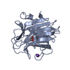 2a6vSC  2a6wC  2a6yC 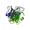 2a6zC  2a70C 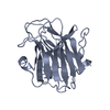 2a71C S: Starting model for refinement C: citing same article ( |
|---|---|
| Similar structure data |

-Links
 PDBj
PDBj- Assembly
| Deposited unit | 
| ||||||||
|---|---|---|---|---|---|---|---|---|---|
| 1 | 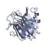
| ||||||||
| 2 | 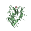
| ||||||||
| Unit cell |
|
-Components
| #1: Protein | Mass: 25577.084 Da / Num. of mol.: 2 / Fragment: RESIDUES 6-229 / Mutation: Y131F Source method: isolated from a genetically manipulated source Source: (gene. exp.) Plasmid: pGEX4T-1 / Species (production host): Escherichia coli / Production host:  #2: Chemical | ChemComp-K / |   Mass: 39.098 Da / Num. of mol.: 1 / Source method: obtained synthetically / Formula: K Mass: 39.098 Da / Num. of mol.: 1 / Source method: obtained synthetically / Formula: K#3: Chemical |   Mass: 62.068 Da / Num. of mol.: 2 / Source method: obtained synthetically / Formula: C2H6O2 Mass: 62.068 Da / Num. of mol.: 2 / Source method: obtained synthetically / Formula: C2H6O2#4: Water | ChemComp-HOH / |  Mass: 18.015 Da / Num. of mol.: 423 / Source method: isolated from a natural source / Formula: H2O Mass: 18.015 Da / Num. of mol.: 423 / Source method: isolated from a natural source / Formula: H2OHas protein modification | Y | |
|---|
-Experimental details
-Experiment
| Experiment | Method: X-RAY DIFFRACTION / Number of used crystals: 1 |
|---|
- Sample preparation
| Crystal | Density Matthews: 2.2 Å3/Da / Density % sol: 43.3 % |
|---|---|
| Crystal grow | Temperature: 289 K / Method: vapor diffusion, hanging drop / pH: 7.5 Details: PEG3350, Potassium fluoride, HEPES, Ethylene glycol, pH 7.5, VAPOR DIFFUSION, HANGING DROP, temperature 289K |
-Data collection
| Diffraction | Mean temperature: 100 K |
|---|---|
| Diffraction source | Source: SYNCHROTRON / Site: Photon Factory  / Beamline: BL-5A / Wavelength: 1 Å / Beamline: BL-5A / Wavelength: 1 Å |
| Detector | Type: ADSC QUANTUM 315 / Detector: CCD / Date: Nov 14, 2004 |
| Radiation | Monochromator: Si(111) / Protocol: SINGLE WAVELENGTH / Monochromatic (M) / Laue (L): M / Scattering type: x-ray |
| Radiation wavelength | Wavelength: 1 Å / Relative weight: 1 |
| Reflection | Resolution: 1.55→50 Å / Num. all: 64255 / Num. obs: 63029 / % possible obs: 98.1 % / Redundancy: 3.6 % / Biso Wilson estimate: 24.6 Å2 / Rmerge(I) obs: 0.053 / Net I/σ(I): 15.6 |
| Reflection shell | Resolution: 1.55→1.61 Å / Redundancy: 3.3 % / Rmerge(I) obs: 0.381 / Mean I/σ(I) obs: 2.8 / Num. unique all: 6196 / % possible all: 97.3 |
- Processing
| Software |
| ||||||||||||||||||||||||||||||||||||||||||||||||||||||||||||||||||||||||||||||||||||||||||||||||||||||||||||||||||||||||||||||||||||||||||||||||||||||||||||||||||||||||||
|---|---|---|---|---|---|---|---|---|---|---|---|---|---|---|---|---|---|---|---|---|---|---|---|---|---|---|---|---|---|---|---|---|---|---|---|---|---|---|---|---|---|---|---|---|---|---|---|---|---|---|---|---|---|---|---|---|---|---|---|---|---|---|---|---|---|---|---|---|---|---|---|---|---|---|---|---|---|---|---|---|---|---|---|---|---|---|---|---|---|---|---|---|---|---|---|---|---|---|---|---|---|---|---|---|---|---|---|---|---|---|---|---|---|---|---|---|---|---|---|---|---|---|---|---|---|---|---|---|---|---|---|---|---|---|---|---|---|---|---|---|---|---|---|---|---|---|---|---|---|---|---|---|---|---|---|---|---|---|---|---|---|---|---|---|---|---|---|---|---|---|---|
| Refinement | Method to determine structure: MOLECULAR REPLACEMENT Starting model: PDB ENTRY 2A6V Resolution: 1.55→20 Å / Cor.coef. Fo:Fc: 0.959 / Cor.coef. Fo:Fc free: 0.945 / SU B: 1.825 / SU ML: 0.066 / Cross valid method: THROUGHOUT / σ(F): 0 / ESU R: 0.095 / ESU R Free: 0.096 / Stereochemistry target values: MAXIMUM LIKELIHOOD
| ||||||||||||||||||||||||||||||||||||||||||||||||||||||||||||||||||||||||||||||||||||||||||||||||||||||||||||||||||||||||||||||||||||||||||||||||||||||||||||||||||||||||||
| Solvent computation | Ion probe radii: 0.8 Å / Shrinkage radii: 0.8 Å / VDW probe radii: 1.4 Å / Solvent model: BABINET MODEL WITH MASK | ||||||||||||||||||||||||||||||||||||||||||||||||||||||||||||||||||||||||||||||||||||||||||||||||||||||||||||||||||||||||||||||||||||||||||||||||||||||||||||||||||||||||||
| Displacement parameters | Biso mean: 23.54 Å2
| ||||||||||||||||||||||||||||||||||||||||||||||||||||||||||||||||||||||||||||||||||||||||||||||||||||||||||||||||||||||||||||||||||||||||||||||||||||||||||||||||||||||||||
| Refinement step | Cycle: LAST / Resolution: 1.55→20 Å
| ||||||||||||||||||||||||||||||||||||||||||||||||||||||||||||||||||||||||||||||||||||||||||||||||||||||||||||||||||||||||||||||||||||||||||||||||||||||||||||||||||||||||||
| Refine LS restraints |
| ||||||||||||||||||||||||||||||||||||||||||||||||||||||||||||||||||||||||||||||||||||||||||||||||||||||||||||||||||||||||||||||||||||||||||||||||||||||||||||||||||||||||||
| LS refinement shell | Highest resolution: 1.55 Å / Num. reflection Rwork: 4335 / Total num. of bins used: 20 |
