 Movie
Movie Controller

[English] 日本語
 Yorodumi
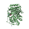
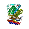
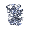
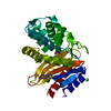
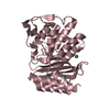
- PDB-5ywv: Crystal structure of TREX1 in complex with a inosine contained ssDNA -
+ Open data

- Basic information
| Entry | Database: PDB / ID: 5ywv | ||||||
|---|---|---|---|---|---|---|---|
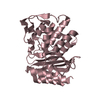
| Title | Crystal structure of TREX1 in complex with a inosine contained ssDNA | ||||||
 Components Components |
| ||||||
 Keywords Keywords | DNA BINDING PROTEIN/DNA / exonuclease / DEDDh family / protein-DNA complex / DNA BINDING PROTEIN-DNA complex | ||||||
| Function / homology |  Function and homology information Function and homology informationcellular response to type I interferon / immune response in brain or nervous system / adenyl deoxyribonucleotide binding / immune complex formation / activation of immune response / organ or tissue specific immune response / DNA synthesis involved in UV-damage excision repair / atrial cardiac muscle tissue development / T cell antigen processing and presentation / MutSalpha complex binding ...cellular response to type I interferon / immune response in brain or nervous system / adenyl deoxyribonucleotide binding / immune complex formation / activation of immune response / organ or tissue specific immune response / DNA synthesis involved in UV-damage excision repair / atrial cardiac muscle tissue development / T cell antigen processing and presentation / MutSalpha complex binding / retrotransposition / DNA exonuclease activity / DNA modification / regulation of lipid biosynthetic process / oligosaccharyltransferase complex / regulation of fatty acid metabolic process / heart process / regulation of protein complex stability / exodeoxyribonuclease III / lymphoid progenitor cell differentiation / double-stranded DNA 3'-5' DNA exonuclease activity / regulation of type I interferon production / regulation of lysosome organization / glycoprotein biosynthetic process / cellular response to hydroxyurea / regulation of tumor necrosis factor production / MutLalpha complex binding / 3'-5'-DNA exonuclease activity / regulation of immunoglobulin production / inflammatory response to antigenic stimulus / macrophage activation involved in immune response / regulation of cellular respiration / DNA catabolic process / regulation of T cell activation / apoptotic cell clearance / regulation of glycolytic process / DNA binding, bending / cGAS/STING signaling pathway / negative regulation of type I interferon-mediated signaling pathway / WW domain binding / regulation of innate immune response / DNA metabolic process / negative regulation of cGAS/STING signaling pathway / type I interferon-mediated signaling pathway / blood vessel development / nuclear replication fork / response to UV / heart morphogenesis / cellular response to interferon-beta / mitotic G1 DNA damage checkpoint signaling / 3'-5' exonuclease activity / negative regulation of innate immune response / DNA damage checkpoint signaling / determination of adult lifespan / generation of precursor metabolites and energy / kidney development / cellular response to reactive oxygen species / establishment of protein localization / cellular response to gamma radiation / protein-DNA complex / cellular response to UV / single-stranded DNA binding / regulation of gene expression / cellular response to oxidative stress / regulation of inflammatory response / double-stranded DNA binding / defense response to virus / adaptive immune response / DNA replication / protein stabilization / immune response / inflammatory response / innate immune response / DNA damage response / endoplasmic reticulum membrane / magnesium ion binding / endoplasmic reticulum / protein homodimerization activity / DNA binding / identical protein binding / nucleus / cytoplasm / cytosol Similarity search - Function | ||||||
| Biological species |   Homo sapiens (human) Homo sapiens (human) | ||||||
| Method |  X-RAY DIFFRACTION / SYNCHROTRON / MOLECULAR REPLACEMENT / Resolution: 2.3 Å X-RAY DIFFRACTION / SYNCHROTRON / MOLECULAR REPLACEMENT / Resolution: 2.3 Å | ||||||
 Authors Authors | Hsiao, Y.Y. | ||||||
| Funding support |  Taiwan, 1items Taiwan, 1items
| ||||||
 Citation Citation | Journal: PLoS Biol. / Year: 2018 Title: Structural basis for overhang excision and terminal unwinding of DNA duplexes by TREX1 Authors: Huang, K.W. / Liu, T.C. / Liang, R.Y. / Chu, L.Y. / Cheng, H.L. / Chu, J.W. / Hsiao, Y.Y. | ||||||
| History |
|
- Structure visualization
| Structure viewer | Molecule: MolmilJmol/JSmol |
|---|
- Downloads & links
-Download
| PDBx/mmCIF format | 5ywv.cif.gz | 208 KB | Display | PDBx/mmCIF format |
|---|---|---|---|---|
| PDB format | pdb5ywv.ent.gz | 161.6 KB | Display | PDB format |
| PDBx/mmJSON format | 5ywv.json.gz | Tree view | PDBx/mmJSON format | |
| Others |  Other downloads Other downloads |
-Validation report
| Arichive directory | https://data.pdbj.org/pub/pdb/validation_reports/yw/5ywvftp://data.pdbj.org/pub/pdb/validation_reports/yw/5ywv | HTTPS FTP |
|---|
-Related structure data
| Related structure data |  5ywsC 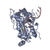 5ywtC 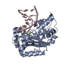 5ywuC 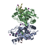 3mxmS S: Starting model for refinement C: citing same article ( |
|---|---|
| Similar structure data |


-Links
 PDBj
PDBj- Assembly
| Deposited unit | 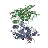
| ||||||||
|---|---|---|---|---|---|---|---|---|---|
| 1 | 
| ||||||||
| 2 | 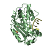
| ||||||||
| Unit cell |
|
-Components
-Protein / DNA chain , 2 types, 4 molecules ABCD
| #1: Protein | Mass: 27987.828 Da / Num. of mol.: 2 Source method: isolated from a genetically manipulated source Source: (gene. exp.)  #2: DNA chain | Mass: 2137.432 Da / Num. of mol.: 2 / Source method: obtained synthetically / Source: (synth.) Homo sapiens (human) |
|---|
-Non-polymers , 5 types, 184 molecules 


| #3: Chemical | ChemComp-MG /  Mass: 24.305 Da / Num. of mol.: 4 / Source method: obtained synthetically / Formula: Mg Mass: 24.305 Da / Num. of mol.: 4 / Source method: obtained synthetically / Formula: Mg#4: Chemical | ChemComp-NA /  Mass: 22.990 Da / Num. of mol.: 4 / Source method: obtained synthetically / Formula: Na Mass: 22.990 Da / Num. of mol.: 4 / Source method: obtained synthetically / Formula: Na#5: Chemical | ChemComp-EDO / |  Mass: 62.068 Da / Num. of mol.: 1 / Source method: obtained synthetically / Formula: C2H6O2 Mass: 62.068 Da / Num. of mol.: 1 / Source method: obtained synthetically / Formula: C2H6O2#6: Chemical | ChemComp-MES / |  Mass: 195.237 Da / Num. of mol.: 1 / Source method: obtained synthetically / Formula: C6H13NO4S / Comment: pH buffer*YM Mass: 195.237 Da / Num. of mol.: 1 / Source method: obtained synthetically / Formula: C6H13NO4S / Comment: pH buffer*YM#7: Water | ChemComp-HOH / | Mass: 18.015 Da / Num. of mol.: 174 / Source method: isolated from a natural source / Formula: H2O |
|---|
-Experimental details
-Experiment
| Experiment | Method: X-RAY DIFFRACTION / Number of used crystals: 1 |
|---|
- Sample preparation
| Crystal | Density Matthews: 1.86 Å3/Da / Density % sol: 33.91 % |
|---|---|
| Crystal grow | Temperature: 293 K / Method: vapor diffusion, hanging drop / pH: 6 Details: 0.1M MES MONOHYDRATE PH 6.0, 20%(W/V) POLYETHYLENE GLYCOL MONOMETHYL ETHER 2,000, VAPOR DIFFUSION, HANGING DROP, TEMPERATURE 293K PH range: 6 |
-Data collection
| Diffraction | Mean temperature: 100 K |
|---|---|
| Diffraction source | Source: SYNCHROTRON / Site: NSRRC / Beamline: BL13B1 / Wavelength: 1 Å |
| Detector | Type: ADSC QUANTUM 315 / Detector: CCD / Date: Nov 3, 2015 |
| Radiation | Protocol: SINGLE WAVELENGTH / Monochromatic (M) / Laue (L): M / Scattering type: x-ray |
| Radiation wavelength | Wavelength: 1 Å / Relative weight: 1 |
| Reflection | Resolution: 2.3→30 Å / Num. obs: 20577 / % possible obs: 99.7 % / Redundancy: 5.9 % / Rmerge(I) obs: 0.107 / Net I/σ(I): 12.8 |
| Reflection shell | Resolution: 2.3→2.38 Å / Redundancy: 5.7 % / Rmerge(I) obs: 0.495 / Mean I/σ(I) obs: 2.25 / % possible all: 100 |
- Processing
| Software |
| ||||||||||||||||||||||||||||||||||||||||||||||||||||||||||||||||||||||||||||||||||||
|---|---|---|---|---|---|---|---|---|---|---|---|---|---|---|---|---|---|---|---|---|---|---|---|---|---|---|---|---|---|---|---|---|---|---|---|---|---|---|---|---|---|---|---|---|---|---|---|---|---|---|---|---|---|---|---|---|---|---|---|---|---|---|---|---|---|---|---|---|---|---|---|---|---|---|---|---|---|---|---|---|---|---|---|---|---|
| Refinement | Method to determine structure: MOLECULAR REPLACEMENT Starting model: 3MXM Resolution: 2.3→29.31 Å / SU ML: 0.22 / Cross valid method: FREE R-VALUE / σ(F): 1.36 / Phase error: 20.13
| ||||||||||||||||||||||||||||||||||||||||||||||||||||||||||||||||||||||||||||||||||||
| Solvent computation | Shrinkage radii: 0.9 Å / VDW probe radii: 1.11 Å | ||||||||||||||||||||||||||||||||||||||||||||||||||||||||||||||||||||||||||||||||||||
| Refinement step | Cycle: LAST / Resolution: 2.3→29.31 Å
| ||||||||||||||||||||||||||||||||||||||||||||||||||||||||||||||||||||||||||||||||||||
| Refine LS restraints |
| ||||||||||||||||||||||||||||||||||||||||||||||||||||||||||||||||||||||||||||||||||||
| LS refinement shell |
| ||||||||||||||||||||||||||||||||||||||||||||||||||||||||||||||||||||||||||||||||||||
| Refinement TLS params. | Method: refined / Origin x: -26.1873 Å / Origin y: 7.7346 Å / Origin z: -21.9613 Å
| ||||||||||||||||||||||||||||||||||||||||||||||||||||||||||||||||||||||||||||||||||||
| Refinement TLS group | Selection details: ALL |
