 Movie
Movie Controller


+ Open data

- Basic information
| Entry | Database: PDB / ID: 5ydt | ||||||
|---|---|---|---|---|---|---|---|
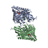
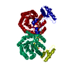
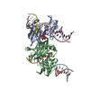
| Title | Remodeled Utp30 in 90S pre-ribosome (Mtr4-depleted, Enp1-TAP) | ||||||
 Components Components |
| ||||||
 Keywords Keywords | RIBOSOME / ribosome assembly / 90S pre-ribosome | ||||||
| Function / homology |  Function and homology information Function and homology information90S preribosome / maturation of SSU-rRNA / maturation of SSU-rRNA from tricistronic rRNA transcript (SSU-rRNA, 5.8S rRNA, LSU-rRNA) / ribosomal small subunit biogenesis / nucleolus / RNA binding / nucleus Similarity search - Function | ||||||
| Biological species |  | ||||||
| Method | ELECTRON MICROSCOPY / single particle reconstruction / cryo EM / Resolution: 4.5 Å | ||||||
 Authors Authors | Ye, K. / Zhu, X. / Hu, J. | ||||||
| Funding support |  China, 1items China, 1items
| ||||||
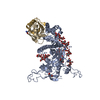
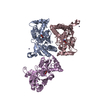
 Citation Citation | Journal: RNA / Year: 2017 Title: Structure and RNA recognition of ribosome assembly factor Utp30. Authors: Jianfei Hu / Xing Zhu / Keqiong Ye / Abstract: The 90S preribosomes are gigantic early assembly intermediates of small ribosomal subunits. Cryo-EM structures of 90S were recently determined, but many of its components have not been accurately ...The 90S preribosomes are gigantic early assembly intermediates of small ribosomal subunits. Cryo-EM structures of 90S were recently determined, but many of its components have not been accurately modeled. Here we determine the crystal structure of yeast Utp30, a ribosomal L1 domain-containing protein in 90S, at 2.65 Å resolution, revealing a classic two-domain fold. The structure of Utp30 fits well into the cryo-EM density of 90S, confirming its previously assigned location. Utp30 binds to the rearranged helix 41 of 18S rRNA and helix 4 of 5' external transcribed spacer in 90S. Comparison of RNA-binding modes of different L1 domains illustrates that they consistently recognize a short RNA duplex with the concaved surface of domain I, but are versatile in RNA recognition outside the core interface. Cic1 is a paralog of Utp30 associating with large subunit preribosomes. Utp30 and Cic1 share similar RNA-binding modes, suggesting that their distinct functions may be executed by a single protein in other organisms. Deletion of Utp30 does not affect the composition of 90S. The nonessential role of Utp30 could be ascribed to its peripheral localization and redundant interactions in 90S. | ||||||
| History |
|
- Structure visualization
| Movie |
Movie viewer |
|---|---|
| Structure viewer | Molecule: MolmilJmol/JSmol |
- Downloads & links
-Download
| PDBx/mmCIF format | 5ydt.cif.gz | 97.7 KB | Display | PDBx/mmCIF format |
|---|---|---|---|---|
| PDB format | pdb5ydt.ent.gz | Display | PDB format | |
| PDBx/mmJSON format | 5ydt.json.gz | Tree view | PDBx/mmJSON format | |
| Others |  Other downloads Other downloads |
-Validation report
| Arichive directory | https://data.pdbj.org/pub/pdb/validation_reports/yd/5ydtftp://data.pdbj.org/pub/pdb/validation_reports/yd/5ydt | HTTPS FTP |
|---|
-Related structure data
| Related structure data | 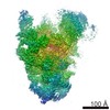 6696M 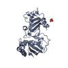 5yduC M: map data used to model this data C: citing same article ( |
|---|---|
| Similar structure data |


-Links
 PDBj
PDBj


- Assembly
| Deposited unit | 
|
|---|---|
| 1 |
|
-Components
| #1: Protein | Mass: 31668.939 Da / Num. of mol.: 1 / Source method: isolated from a natural source Source: (natural) Strain: ATCC 204508 / S288c / References: UniProt: P36144 |
|---|---|
| #2: RNA chain | Mass: 20614.229 Da / Num. of mol.: 1 / Source method: isolated from a natural source / Source: (natural) |
| #3: RNA chain | Mass: 6031.593 Da / Num. of mol.: 1 / Source method: isolated from a natural source / Source: (natural) |
| #4: Protein/peptide | Mass: 2911.580 Da / Num. of mol.: 1 / Source method: isolated from a natural source / Source: (natural) |
-Experimental details
-Experiment
| Experiment | Method: ELECTRON MICROSCOPY |
|---|---|
| EM experiment | Aggregation state: PARTICLE / 3D reconstruction method: single particle reconstruction |
- Sample preparation
| Component | Name: 90S small subunit pre-ribosome (Mtr4-depleted, Enp1-TAP) Type: RIBOSOME / Entity ID: all / Source: NATURAL |
|---|---|
| Molecular weight | Experimental value: NO |
| Source (natural) | Organism: |
| Buffer solution | pH: 8 |
| Specimen | Embedding applied: NO / Shadowing applied: NO / Staining applied: NO / Vitrification applied: YES |
| Vitrification | Cryogen name: ETHANE / Humidity: 100 % / Chamber temperature: 277.15 K |
- Electron microscopy imaging
| Experimental equipment |  Model: Titan Krios / Image courtesy: FEI Company |
|---|---|
| Microscopy | Model: FEI TITAN KRIOS |
| Electron gun | Electron source:  FIELD EMISSION GUN / Accelerating voltage: 300 kV / Illumination mode: FLOOD BEAM FIELD EMISSION GUN / Accelerating voltage: 300 kV / Illumination mode: FLOOD BEAM |
| Electron lens | Mode: BRIGHT FIELD |
| Image recording | Electron dose: 40 e/Å2 / Film or detector model: FEI FALCON III (4k x 4k) |
- Processing
| EM software | Name: RELION / Version: 1.4 / Category: 3D reconstruction |
|---|---|
| CTF correction | Type: PHASE FLIPPING ONLY |
| Symmetry | Point symmetry: C1 (asymmetric) |
| 3D reconstruction | Resolution: 4.5 Å / Resolution method: FSC 0.143 CUT-OFF / Num. of particles: 73543 / Symmetry type: POINT |
| Atomic model building | Space: REAL |
