RIBOSOMAL PROTEIN / ribosome assembly / 90S pre-ribosome
Function / homology
Function and homology information
90S preribosome / maturation of SSU-rRNA from tricistronic rRNA transcript (SSU-rRNA, 5.8S rRNA, LSU-rRNA) / maturation of SSU-rRNA / ribosomal small subunit biogenesis / nucleolus / RNA binding / nucleus Similarity search - Function
Ribosomal protein L1-like / Ribosomal protein L1/ribosomal biogenesis protein / Ribosomal protein L1p/L10e family / : Similarity search - Domain/homology
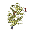
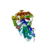
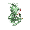
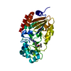
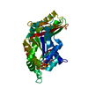
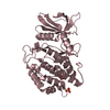
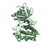
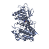
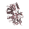
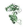
Journal: RNA / Year: 2017 Title: Structure and RNA recognition of ribosome assembly factor Utp30. Authors: Jianfei Hu / Xing Zhu / Keqiong Ye / Abstract: The 90S preribosomes are gigantic early assembly intermediates of small ribosomal subunits. Cryo-EM structures of 90S were recently determined, but many of its components have not been accurately ...The 90S preribosomes are gigantic early assembly intermediates of small ribosomal subunits. Cryo-EM structures of 90S were recently determined, but many of its components have not been accurately modeled. Here we determine the crystal structure of yeast Utp30, a ribosomal L1 domain-containing protein in 90S, at 2.65 Å resolution, revealing a classic two-domain fold. The structure of Utp30 fits well into the cryo-EM density of 90S, confirming its previously assigned location. Utp30 binds to the rearranged helix 41 of 18S rRNA and helix 4 of 5' external transcribed spacer in 90S. Comparison of RNA-binding modes of different L1 domains illustrates that they consistently recognize a short RNA duplex with the concaved surface of domain I, but are versatile in RNA recognition outside the core interface. Cic1 is a paralog of Utp30 associating with large subunit preribosomes. Utp30 and Cic1 share similar RNA-binding modes, suggesting that their distinct functions may be executed by a single protein in other organisms. Deletion of Utp30 does not affect the composition of 90S. The nonessential role of Utp30 could be ascribed to its peripheral localization and redundant interactions in 90S.
In the structure databanks used in Yorodumi, some data are registered as the other names, "COVID-19 virus" and "2019-nCoV". Here are the details of the virus and the list of structure data.
Jan 31, 2019. EMDB accession codes are about to change! (news from PDBe EMDB page)
EMDB accession codes are about to change! (news from PDBe EMDB page)
The allocation of 4 digits for EMDB accession codes will soon come to an end. Whilst these codes will remain in use, new EMDB accession codes will include an additional digit and will expand incrementally as the available range of codes is exhausted. The current 4-digit format prefixed with “EMD-” (i.e. EMD-XXXX) will advance to a 5-digit format (i.e. EMD-XXXXX), and so on. It is currently estimated that the 4-digit codes will be depleted around Spring 2019, at which point the 5-digit format will come into force.
The EM Navigator/Yorodumi systems omit the EMD- prefix.
Related info.:Q: What is EMD? / ID/Accession-code notation in Yorodumi/EM Navigator
Yorodumi is a browser for structure data from EMDB, PDB, SASBDB, etc.
This page is also the successor to EM Navigator detail page, and also detail information page/front-end page for Omokage search.
The word "yorodu" (or yorozu) is an old Japanese word meaning "ten thousand". "mi" (miru) is to see.
Related info.:EMDB / PDB / SASBDB / Comparison of 3 databanks / Yorodumi Search / Aug 31, 2016. New EM Navigator & Yorodumi / Yorodumi Papers / Jmol/JSmol / Function and homology information / Changes in new EM Navigator and Yorodumi
 Movie
Movie Controller


 Open data
 Basic information
 Components
 Keywords
 Function and homology information
 X-RAY DIFFRACTION /
  Authors
 Citation
 Structure visualization
 Downloads & links
 Other downloads
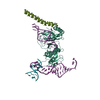

 PDBj
PDBj

 Assembly
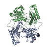


 Mass: 94.971 Da / Num. of mol.: 1 / Source method: obtained synthetically / Formula: PO4
 Mass: 18.015 Da / Num. of mol.: 15 / Source method: isolated from a natural source / Formula: H2O
 Sample preparation
 Processing