 Movie
Movie Controller

[English] 日本語
 Yorodumi
- PDB-5fm4: Structure of the C-terminally extended domain My4 of human myomes... -
+ Open data

- Basic information
| Entry | Database: PDB / ID: 5fm4 | |||||||||
|---|---|---|---|---|---|---|---|---|---|---|
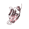
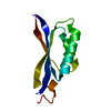
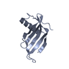
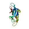
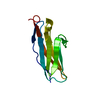
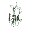
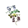
| Title | Structure of the C-terminally extended domain My4 of human myomesin (space group P21) | |||||||||
 Components Components | MYOMESIN-1 | |||||||||
 Keywords Keywords | STRUCTURAL PROTEIN / SARCOMERE / M-BAND / FIBRONECTIN DOMAIN | |||||||||
| Function / homology |  Function and homology information Function and homology informationextraocular skeletal muscle development / striated muscle myosin thick filament / M band / sarcomere organization / positive regulation of cAMP/PKA signal transduction / structural constituent of muscle / kinase binding / positive regulation of gene expression / protein homodimerization activity / identical protein binding Similarity search - Function | |||||||||
| Biological species |  HOMO SAPIENS (human) HOMO SAPIENS (human) | |||||||||
| Method |  X-RAY DIFFRACTION / SYNCHROTRON / MOLECULAR REPLACEMENT / Resolution: 2.8 Å X-RAY DIFFRACTION / SYNCHROTRON / MOLECULAR REPLACEMENT / Resolution: 2.8 Å | |||||||||
 Authors Authors | Pernigo, S. / Steiner, R.A. | |||||||||
 Citation Citation | Journal: Structure / Year: 2017 Title: Binding of Myomesin to Obscurin-Like-1 at the Muscle M-Band Provides a Strategy for Isoform-Specific Mechanical Protection. Authors: Pernigo, S. / Fukuzawa, A. / Beedle, A.E. / Holt, M. / Round, A. / Pandini, A. / Garcia-Manyes, S. / Gautel, M. / Steiner, R.A. | |||||||||
| History |
|
- Structure visualization
| Structure viewer | Molecule: MolmilJmol/JSmol |
|---|
- Downloads & links
-Download
| PDBx/mmCIF format | 5fm4.cif.gz | 223.2 KB | Display | PDBx/mmCIF format |
|---|---|---|---|---|
| PDB format | pdb5fm4.ent.gz | 184.5 KB | Display | PDB format |
| PDBx/mmJSON format | 5fm4.json.gz | Tree view | PDBx/mmJSON format | |
| Others |  Other downloads Other downloads |
-Validation report
| Arichive directory | https://data.pdbj.org/pub/pdb/validation_reports/fm/5fm4ftp://data.pdbj.org/pub/pdb/validation_reports/fm/5fm4 | HTTPS FTP |
|---|
-Related structure data
| Related structure data | 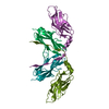 5fm5SC 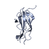 5fm8C S: Starting model for refinement C: citing same article ( |
|---|---|
| Similar structure data |


-Links
 PDBj
PDBj


- Assembly
| Deposited unit | 
| ||||||||
|---|---|---|---|---|---|---|---|---|---|
| 1 | 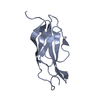
| ||||||||
| 2 | 
| ||||||||
| 3 | 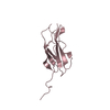
| ||||||||
| 4 | 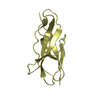
| ||||||||
| 5 | 
| ||||||||
| 6 | 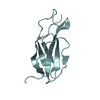
| ||||||||
| Unit cell |
|
-Components
| #1: Protein | Mass: 12186.966 Da / Num. of mol.: 6 Fragment: MY4 EXTENDED AT ITS C-TERMINUS, UNP RESIDUES 510-618 Source method: isolated from a genetically manipulated source Source: (gene. exp.) HOMO SAPIENS (human) / Production host:  #2: Water | ChemComp-HOH / |  Mass: 18.015 Da / Num. of mol.: 37 / Source method: isolated from a natural source / Formula: H2O Mass: 18.015 Da / Num. of mol.: 37 / Source method: isolated from a natural source / Formula: H2O |
|---|
-Experimental details
-Experiment
| Experiment | Method: X-RAY DIFFRACTION / Number of used crystals: 1 |
|---|
- Sample preparation
| Crystal | Density Matthews: 2.25 Å3/Da / Density % sol: 45.3 % / Description: NONE |
|---|---|
| Crystal grow | pH: 5.5 / Details: 25 % (W/V) PEG 3350 100 MM BIS-TRIS PH 5.5 |
-Data collection
| Diffraction | Mean temperature: 100 K |
|---|---|
| Diffraction source | Source: SYNCHROTRON / Site: Diamond  / Beamline: I04-1 / Wavelength: 0.92819 / Beamline: I04-1 / Wavelength: 0.92819 |
| Detector | Type: DECTRIS PILATUS 6M / Detector: PIXEL / Date: Sep 25, 2015 |
| Radiation | Protocol: SINGLE WAVELENGTH / Monochromatic (M) / Laue (L): M / Scattering type: x-ray |
| Radiation wavelength | Wavelength: 0.92819 Å / Relative weight: 1 |
| Reflection | Resolution: 2.8→39.21 Å / Num. obs: 15138 / % possible obs: 95.3 % / Observed criterion σ(I): -1 / Redundancy: 3.1 % / Biso Wilson estimate: 38.45 Å2 / Rmerge(I) obs: 0.09 / Net I/σ(I): 10.8 |
| Reflection shell | Resolution: 2.8→2.87 Å / Redundancy: 3.1 % / Rmerge(I) obs: 0.31 / Mean I/σ(I) obs: 3.1 / % possible all: 89.8 |
- Processing
| Software |
| |||||||||||||||||||||||||||||||||||||||||||||||||||||||||||||||||||||||||||||
|---|---|---|---|---|---|---|---|---|---|---|---|---|---|---|---|---|---|---|---|---|---|---|---|---|---|---|---|---|---|---|---|---|---|---|---|---|---|---|---|---|---|---|---|---|---|---|---|---|---|---|---|---|---|---|---|---|---|---|---|---|---|---|---|---|---|---|---|---|---|---|---|---|---|---|---|---|---|---|
| Refinement | Method to determine structure: MOLECULAR REPLACEMENT Starting model: PDB ENTRY 5FM5 Resolution: 2.8→39.21 Å / SU ML: 0.4 / σ(F): 0.02 / Phase error: 31.62 / Stereochemistry target values: ML
| |||||||||||||||||||||||||||||||||||||||||||||||||||||||||||||||||||||||||||||
| Solvent computation | Shrinkage radii: 0.9 Å / VDW probe radii: 1.11 Å / Solvent model: FLAT BULK SOLVENT MODEL | |||||||||||||||||||||||||||||||||||||||||||||||||||||||||||||||||||||||||||||
| Displacement parameters | Biso mean: 35.3 Å2 | |||||||||||||||||||||||||||||||||||||||||||||||||||||||||||||||||||||||||||||
| Refinement step | Cycle: LAST / Resolution: 2.8→39.21 Å
| |||||||||||||||||||||||||||||||||||||||||||||||||||||||||||||||||||||||||||||
| Refine LS restraints |
| |||||||||||||||||||||||||||||||||||||||||||||||||||||||||||||||||||||||||||||
| LS refinement shell |
|
