 Movie
Movie Controller

[English] 日本語
 Yorodumi
- PDB-5ybu: Structure of the KANK1 ankyrin domain in complex with KIF21A peptide -
+ Open data

- Basic information
| Entry | Database: PDB / ID: 5ybu | ||||||
|---|---|---|---|---|---|---|---|
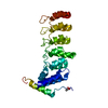
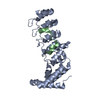
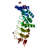
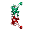
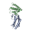
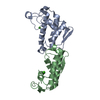
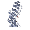
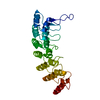
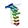
| Title | Structure of the KANK1 ankyrin domain in complex with KIF21A peptide | ||||||
 Components Components |
| ||||||
 Keywords Keywords | CELL ADHESION | ||||||
| Function / homology |  Function and homology information Function and homology informationregulation of microtubule depolymerization / negative regulation of lamellipodium morphogenesis / negative regulation of ruffle assembly / regulation of axon guidance / podocyte cell migration / negative regulation of substrate adhesion-dependent cell spreading / Signaling by membrane-tethered fusions of PDGFRA or PDGFRB / negative regulation of actin filament polymerization / cortical microtubule organization / ankyrin repeat binding ...regulation of microtubule depolymerization / negative regulation of lamellipodium morphogenesis / negative regulation of ruffle assembly / regulation of axon guidance / podocyte cell migration / negative regulation of substrate adhesion-dependent cell spreading / Signaling by membrane-tethered fusions of PDGFRA or PDGFRB / negative regulation of actin filament polymerization / cortical microtubule organization / ankyrin repeat binding / regulation of Rho protein signal transduction / anterograde axonal transport / plus-end-directed microtubule motor activity / Kinesins / negative regulation of Rho protein signal transduction / microtubule motor activity / kinesin complex / COPI-dependent Golgi-to-ER retrograde traffic / microtubule-based movement / positive regulation of wound healing / regulation of establishment of cell polarity / regulation of microtubule polymerization / positive regulation of Wnt signaling pathway / axonal growth cone / axon cytoplasm / negative regulation of insulin receptor signaling pathway / negative regulation of cell migration / beta-catenin binding / ruffle membrane / positive regulation of canonical Wnt signaling pathway / negative regulation of neuron projection development / presynapse / actin cytoskeleton organization / cell cortex / Estrogen-dependent gene expression / microtubule binding / microtubule / cytoskeleton / protein-macromolecule adaptor activity / cell population proliferation / dendrite / DNA-templated transcription / ATP hydrolysis activity / ATP binding / nucleus / plasma membrane / cytosol / cytoplasm Similarity search - Function | ||||||
| Biological species |  Homo sapiens (human) Homo sapiens (human) | ||||||
| Method |  X-RAY DIFFRACTION / SYNCHROTRON / MOLECULAR REPLACEMENT / Resolution: 1.89 Å X-RAY DIFFRACTION / SYNCHROTRON / MOLECULAR REPLACEMENT / Resolution: 1.89 Å | ||||||
 Authors Authors | Guo, Q. / Liao, S. / Min, J. / Xu, C. / Structural Genomics Consortium (SGC) | ||||||
 Citation Citation | Journal: J. Biol. Chem. / Year: 2018 Title: Structural basis for the recognition of kinesin family member 21A (KIF21A) by the ankyrin domains of KANK1 and KANK2 proteins. Authors: Guo, Q. / Liao, S. / Zhu, Z. / Li, Y. / Li, F. / Xu, C. | ||||||
| History |
|
- Structure visualization
| Structure viewer | Molecule: MolmilJmol/JSmol |
|---|
- Downloads & links
-Download
| PDBx/mmCIF format | 5ybu.cif.gz | 116.8 KB | Display | PDBx/mmCIF format |
|---|---|---|---|---|
| PDB format | pdb5ybu.ent.gz | 89.4 KB | Display | PDB format |
| PDBx/mmJSON format | 5ybu.json.gz | Tree view | PDBx/mmJSON format | |
| Others |  Other downloads Other downloads |
-Validation report
| Arichive directory | https://data.pdbj.org/pub/pdb/validation_reports/yb/5ybuftp://data.pdbj.org/pub/pdb/validation_reports/yb/5ybu | HTTPS FTP |
|---|
-Related structure data
| Related structure data |  5ybjC 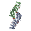 5ybvC 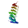 4hbdS S: Starting model for refinement C: citing same article ( |
|---|---|
| Similar structure data |

-Links
 PDBj
PDBj


- Assembly
| Deposited unit | 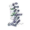
| ||||||||
|---|---|---|---|---|---|---|---|---|---|
| 1 |
| ||||||||
| Unit cell |
|
-Components
| #1: Protein | Mass: 27310.148 Da / Num. of mol.: 1 / Fragment: UNP residues 1080-1329 Source method: isolated from a genetically manipulated source Source: (gene. exp.) Homo sapiens (human) / Gene: KANK1, ANKRD15, KANK, KIAA0172Production host: References: UniProt: Q14678 |
|---|---|
| #2: Protein/peptide | Mass: 2655.081 Da / Num. of mol.: 1 / Fragment: UNP residues 1146-1167 Source method: isolated from a genetically manipulated source Details: KIF21A (1146-1167) / Source: (gene. exp.) Homo sapiens (human) / Gene: KIF21A, KIAA1708, KIF2Production host: References: UniProt: Q7Z4S6 |
| #3: Water | ChemComp-HOH /  Mass: 18.015 Da / Num. of mol.: 95 / Source method: isolated from a natural source / Formula: H2O Mass: 18.015 Da / Num. of mol.: 95 / Source method: isolated from a natural source / Formula: H2O |
-Experimental details
-Experiment
| Experiment | Method: X-RAY DIFFRACTION / Number of used crystals: 1 |
|---|
- Sample preparation
| Crystal | Density Matthews: 2.23 Å3/Da / Density % sol: 44.88 % |
|---|---|
| Crystal grow | Temperature: 291 K / Method: vapor diffusion, sitting drop / Details: 0.05M magnesium formate, 21% PEG 3350 |
-Data collection
| Diffraction | Mean temperature: 100 K |
|---|---|
| Diffraction source | Source: SYNCHROTRON / Site: SSRF  / Beamline: BL17U1 / Wavelength: 0.9796 Å / Beamline: BL17U1 / Wavelength: 0.9796 Å |
| Detector | Type: ADSC QUANTUM 315r / Detector: CCD / Date: Jan 7, 2017 |
| Radiation | Protocol: SINGLE WAVELENGTH / Monochromatic (M) / Laue (L): M / Scattering type: x-ray |
| Radiation wavelength | Wavelength: 0.9796 Å / Relative weight: 1 |
| Reflection | Resolution: 1.89→48.335 Å / Num. obs: 22149 / % possible obs: 99.5 % / Redundancy: 13.9 % / Net I/σ(I): 29.3 |
- Processing
| Software |
| |||||||||||||||||||||||||||||||||||||||||||||||||||||||||||||||||||||||||||||||||||||||||||||||||||||||||||||||||||||||||||||||||||||||||||||||||||||||||||||||||||||||||||||||||||||||||||||||||||||||||||||||||||||||||||||||||
|---|---|---|---|---|---|---|---|---|---|---|---|---|---|---|---|---|---|---|---|---|---|---|---|---|---|---|---|---|---|---|---|---|---|---|---|---|---|---|---|---|---|---|---|---|---|---|---|---|---|---|---|---|---|---|---|---|---|---|---|---|---|---|---|---|---|---|---|---|---|---|---|---|---|---|---|---|---|---|---|---|---|---|---|---|---|---|---|---|---|---|---|---|---|---|---|---|---|---|---|---|---|---|---|---|---|---|---|---|---|---|---|---|---|---|---|---|---|---|---|---|---|---|---|---|---|---|---|---|---|---|---|---|---|---|---|---|---|---|---|---|---|---|---|---|---|---|---|---|---|---|---|---|---|---|---|---|---|---|---|---|---|---|---|---|---|---|---|---|---|---|---|---|---|---|---|---|---|---|---|---|---|---|---|---|---|---|---|---|---|---|---|---|---|---|---|---|---|---|---|---|---|---|---|---|---|---|---|---|---|---|---|---|---|---|---|---|---|---|---|---|---|---|---|---|---|---|
| Refinement | Method to determine structure: MOLECULAR REPLACEMENT Starting model: 4HBD Resolution: 1.89→48.335 Å / Cross valid method: FREE R-VALUE / σ(F): 1.35
| |||||||||||||||||||||||||||||||||||||||||||||||||||||||||||||||||||||||||||||||||||||||||||||||||||||||||||||||||||||||||||||||||||||||||||||||||||||||||||||||||||||||||||||||||||||||||||||||||||||||||||||||||||||||||||||||||
| Solvent computation | Shrinkage radii: 0.9 Å / VDW probe radii: 1.11 Å | |||||||||||||||||||||||||||||||||||||||||||||||||||||||||||||||||||||||||||||||||||||||||||||||||||||||||||||||||||||||||||||||||||||||||||||||||||||||||||||||||||||||||||||||||||||||||||||||||||||||||||||||||||||||||||||||||
| Refinement step | Cycle: LAST / Resolution: 1.89→48.335 Å
| |||||||||||||||||||||||||||||||||||||||||||||||||||||||||||||||||||||||||||||||||||||||||||||||||||||||||||||||||||||||||||||||||||||||||||||||||||||||||||||||||||||||||||||||||||||||||||||||||||||||||||||||||||||||||||||||||
| Refine LS restraints |
| |||||||||||||||||||||||||||||||||||||||||||||||||||||||||||||||||||||||||||||||||||||||||||||||||||||||||||||||||||||||||||||||||||||||||||||||||||||||||||||||||||||||||||||||||||||||||||||||||||||||||||||||||||||||||||||||||
| LS refinement shell |
| |||||||||||||||||||||||||||||||||||||||||||||||||||||||||||||||||||||||||||||||||||||||||||||||||||||||||||||||||||||||||||||||||||||||||||||||||||||||||||||||||||||||||||||||||||||||||||||||||||||||||||||||||||||||||||||||||
| Refinement TLS params. | Method: refined / Refine-ID: X-RAY DIFFRACTION
| |||||||||||||||||||||||||||||||||||||||||||||||||||||||||||||||||||||||||||||||||||||||||||||||||||||||||||||||||||||||||||||||||||||||||||||||||||||||||||||||||||||||||||||||||||||||||||||||||||||||||||||||||||||||||||||||||
| Refinement TLS group |
|
