 Movie
Movie Controller


+ Open data

- Basic information
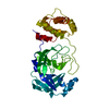
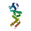
| Entry | Database: PDB / ID: 6tlh | ||||||||||||
|---|---|---|---|---|---|---|---|---|---|---|---|---|---|
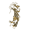
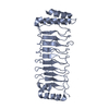
| Title | Crystal structure of mouse KANK3 ankyrin repeats | ||||||||||||
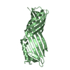
 Components Components | KN motif and ankyrin repeat domain-containing protein 3 | ||||||||||||
 Keywords Keywords | CELL ADHESION / Protein binding / Ankyrin repeat domain-containing protein 47 / actin polymerization | ||||||||||||
| Function / homology |  Function and homology information Function and homology informationnegative regulation of actin filament polymerization / negative regulation of stress fiber assembly / cytoskeleton / cytoplasm Similarity search - Function | ||||||||||||
| Biological species |  | ||||||||||||
| Method |  X-RAY DIFFRACTION / SYNCHROTRON / MOLECULAR REPLACEMENT / Resolution: 1.8 Å X-RAY DIFFRACTION / SYNCHROTRON / MOLECULAR REPLACEMENT / Resolution: 1.8 Å | ||||||||||||
 Authors Authors | Khan, R. / Singh, A.K. / Goult, B.T. | ||||||||||||
| Funding support |  United Kingdom, 3items United Kingdom, 3items
| ||||||||||||
 Citation Citation | Journal: To be published Title: Crystal structure of mouse KANK3 ankyrin repeats Authors: Khan, R. / Singh, A.K. / Goult, B.T. | ||||||||||||
| History |
|
- Structure visualization
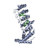
| Structure viewer | Molecule: MolmilJmol/JSmol |
|---|
- Downloads & links
-Download
| PDBx/mmCIF format | 6tlh.cif.gz | 109.9 KB | Display | PDBx/mmCIF format |
|---|---|---|---|---|
| PDB format | pdb6tlh.ent.gz | 84.1 KB | Display | PDB format |
| PDBx/mmJSON format | 6tlh.json.gz | Tree view | PDBx/mmJSON format | |
| Others |  Other downloads Other downloads |
-Validation report
| Arichive directory | https://data.pdbj.org/pub/pdb/validation_reports/tl/6tlhftp://data.pdbj.org/pub/pdb/validation_reports/tl/6tlh | HTTPS FTP |
|---|
-Related structure data
| Related structure data | 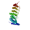 4hbdS S: Starting model for refinement |
|---|---|
| Similar structure data |
-Links
 PDBj
PDBj

- Assembly
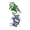
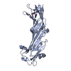
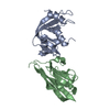
| Deposited unit | 
| |||||||||||||||||||||||||||
|---|---|---|---|---|---|---|---|---|---|---|---|---|---|---|---|---|---|---|---|---|---|---|---|---|---|---|---|---|
| 1 | 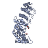
| |||||||||||||||||||||||||||
| 2 | 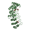
| |||||||||||||||||||||||||||
| Unit cell |
| |||||||||||||||||||||||||||
| Components on special symmetry positions |
| |||||||||||||||||||||||||||
| Noncrystallographic symmetry (NCS) | NCS domain:
NCS domain segments:
|
-Components
| #1: Protein | Mass: 27045.314 Da / Num. of mol.: 2 Source method: isolated from a genetically manipulated source Details: Residues 524-773 / Source: (gene. exp.)  #2: Chemical | ChemComp-GOL /   Mass: 92.094 Da / Num. of mol.: 5 / Source method: obtained synthetically / Formula: C3H8O3 Mass: 92.094 Da / Num. of mol.: 5 / Source method: obtained synthetically / Formula: C3H8O3#3: Chemical | ChemComp-FMT / |   Mass: 46.025 Da / Num. of mol.: 1 / Source method: obtained synthetically / Formula: CH2O2 Mass: 46.025 Da / Num. of mol.: 1 / Source method: obtained synthetically / Formula: CH2O2#4: Water | ChemComp-HOH / |  Mass: 18.015 Da / Num. of mol.: 370 / Source method: isolated from a natural source / Formula: H2O Mass: 18.015 Da / Num. of mol.: 370 / Source method: isolated from a natural source / Formula: H2OHas ligand of interest | N | |
|---|
-Experimental details
-Experiment
| Experiment | Method: X-RAY DIFFRACTION / Number of used crystals: 1 |
|---|
- Sample preparation
| Crystal | Density Matthews: 2.19 Å3/Da / Density % sol: 43.97 % |
|---|---|
| Crystal grow | Temperature: 294 K / Method: vapor diffusion, hanging drop / Details: 0.2 M ammonium formate, 20% w/v PEG 3350 |
-Data collection
| Diffraction | Mean temperature: 100 K / Serial crystal experiment: N |
|---|---|
| Diffraction source | Source: SYNCHROTRON / Site: Diamond / Beamline: I04-1 / Wavelength: 0.91587 Å |
| Detector | Type: DECTRIS PILATUS 6M-F / Detector: PIXEL / Date: Jun 28, 2019 |
| Radiation | Protocol: SINGLE WAVELENGTH / Monochromatic (M) / Laue (L): M / Scattering type: x-ray |
| Radiation wavelength | Wavelength: 0.91587 Å / Relative weight: 1 |
| Reflection | Resolution: 1.8→69.32 Å / Num. obs: 42604 / % possible obs: 97.4 % / Redundancy: 3.5 % / CC1/2: 0.999 / Rmerge(I) obs: 0.062 / Rpim(I) all: 0.039 / Rrim(I) all: 0.073 / Net I/σ(I): 10.5 |
| Reflection shell | Resolution: 1.8→1.84 Å / Redundancy: 3.7 % / Rmerge(I) obs: 0.776 / Mean I/σ(I) obs: 1.5 / Num. unique obs: 2516 / CC1/2: 0.811 / Rpim(I) all: 0.472 / Rrim(I) all: 0.909 / % possible all: 96.7 |
- Processing
| Software |
| ||||||||||||||||||||||||||||||||||||||||||||||||||||||||||||
|---|---|---|---|---|---|---|---|---|---|---|---|---|---|---|---|---|---|---|---|---|---|---|---|---|---|---|---|---|---|---|---|---|---|---|---|---|---|---|---|---|---|---|---|---|---|---|---|---|---|---|---|---|---|---|---|---|---|---|---|---|---|
| Refinement | Method to determine structure: MOLECULAR REPLACEMENT Starting model: 4hbd Resolution: 1.8→53.17 Å / Cor.coef. Fo:Fc: 0.968 / Cor.coef. Fo:Fc free: 0.957 / SU B: 3.749 / SU ML: 0.109 / Cross valid method: THROUGHOUT / σ(F): 0 / ESU R: 0.148 / ESU R Free: 0.137 Details: HYDROGENS HAVE BEEN ADDED IN THE RIDING POSITIONS U VALUES : REFINED INDIVIDUALLY
| ||||||||||||||||||||||||||||||||||||||||||||||||||||||||||||
| Solvent computation | Ion probe radii: 0.8 Å / Shrinkage radii: 0.8 Å / VDW probe radii: 1.2 Å | ||||||||||||||||||||||||||||||||||||||||||||||||||||||||||||
| Displacement parameters | Biso max: 130.28 Å2 / Biso mean: 32.084 Å2 / Biso min: 16.22 Å2
| ||||||||||||||||||||||||||||||||||||||||||||||||||||||||||||
| Refinement step | Cycle: final / Resolution: 1.8→53.17 Å
| ||||||||||||||||||||||||||||||||||||||||||||||||||||||||||||
| Refine LS restraints |
| ||||||||||||||||||||||||||||||||||||||||||||||||||||||||||||
| Refine LS restraints NCS | Number: 1761 / Type: TIGHT THERMAL / Rms dev position: 4.13 Å / Weight position: 0.5 | ||||||||||||||||||||||||||||||||||||||||||||||||||||||||||||
| LS refinement shell | Resolution: 1.8→1.847 Å / Rfactor Rfree error: 0 / Total num. of bins used: 20
|
