Mass: 18.015 Da / Num. of mol.: 11 / Source method: isolated from a natural source / Formula: H2O
Has protein modification
Y
Nonpolymer details
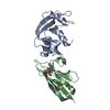
THE ELECTRON DENSITY SUGGESTS THE MODIFICATION OF THE SULFHYDRYL GROUPS OF CYS-1565 AND CYS-1592, ...THE ELECTRON DENSITY SUGGESTS THE MODIFICATION OF THE SULFHYDRYL GROUPS OF CYS-1565 AND CYS-1592, POSSIBLY BY A REACTION INVOLVING CACODYLATE, SIMILAR TO SCOTT ET AL (1993, CHEM RES TOXICOL 6:102) AND GOLDGUR ET AL (1998, PROC NATL ACAD SCI USA 95:9150). ONLY THE PUTATIVE ARSENIC ATOMS OF THE MODIFICATION HAVE BEEN MODELED.
Sequence details
SOLVENT CONTENT AND MATTHEWS COEFFICIENT WERE CALCULATED BASED ON THE PURIFIED PROTEIN CONSTRUCT. ...SOLVENT CONTENT AND MATTHEWS COEFFICIENT WERE CALCULATED BASED ON THE PURIFIED PROTEIN CONSTRUCT. HOWEVER, THE PROTEASE ADDED FOR CRYSTALLIZATION MAY HAVE REDUCED THE CHAIN LENGTH OF THE TARGET PROTEIN.
-
Experimental details
-
Experiment
Experiment
Method: X-RAY DIFFRACTION / Number of used crystals: 1
-
Sample preparation
Crystal
Density Matthews: 2.3 Å3/Da / Density % sol: 46.3 %
Crystal grow
Temperature: 298 K / Method: vapor diffusion, sitting drop / pH: 5.2 Details: 1.39M sodium citrate, 0.1M sodium cacodylate. The protein stock solution was adjusted to contain 15 mM TCEP, supplemented with 1:100 (w/w) chymotrypsin, pH 5.2, VAPOR DIFFUSION, SITTING DROP, temperature 298K
Protocol: SINGLE WAVELENGTH / Monochromatic (M) / Laue (L): M / Scattering type: x-ray
Radiation wavelength
Wavelength: 0.97942 Å / Relative weight: 1
Reflection
Resolution: 2→30 Å / Num. obs: 7245 / % possible obs: 99.5 % / Redundancy: 4.4 % / Rmerge(I) obs: 0.086 / Χ2: 2.407 / Net I/σ(I): 9.6
Reflection shell
Resolution (Å)
Redundancy (%)
Rmerge(I) obs
Num. unique all
Χ2
% possible all
2-2.03
3.6
0.499
381
1.34
99.7
2.03-2.07
3.6
0.553
343
2.671
99.4
2.07-2.11
4.1
0.446
357
1.21
100
2.11-2.15
4.2
0.416
356
1.252
99.7
2.15-2.2
4.4
0.329
365
1.173
100
2.2-2.25
4.4
0.383
355
1.44
98.3
2.25-2.31
4.6
0.3
333
1.597
100
2.31-2.37
4.7
0.281
378
1.247
100
2.37-2.44
4.7
0.208
363
1.173
100
2.44-2.52
4.7
0.191
360
1.466
99.7
2.52-2.61
4.8
0.173
353
1.411
100
2.61-2.71
4.7
0.144
376
1.524
99.7
2.71-2.84
4.8
0.119
348
1.824
100
2.84-2.99
4.7
0.102
364
2.051
99.7
2.99-3.17
4.6
0.094
353
2.362
100
3.17-3.42
4.6
0.069
371
2.534
99.2
3.42-3.76
4.6
0.068
366
3.242
98.7
3.76-4.31
4.4
0.063
356
4.2
98.1
4.31-5.42
4.2
0.055
383
4.443
99.2
5.42-30
3.9
0.069
384
10.945
98.5
-
Phasing
Phasing
Method: SAD
-
Processing
Software
Name
Version
Classification
NB
DENZO
datareduction
SCALEPACK
datascaling
SHELX
phasing
DM
phasing
REFMAC
5.5.0072
refinement
PDB_EXTRACT
3.005
dataextraction
Refinement
Method to determine structure: SAD / Resolution: 2.004→30 Å / Cor.coef. Fo:Fc: 0.935 / Cor.coef. Fo:Fc free: 0.887 / WRfactor Rfree: 0.27 / WRfactor Rwork: 0.239 / SU B: 11.569 / SU ML: 0.154 / Cross valid method: THROUGHOUT / σ(F): 0 / ESU R: 0.228 / ESU R Free: 0.191 / Stereochemistry target values: MAXIMUM LIKELIHOOD Details: HYDROGENS HAVE BEEN ADDED IN THE RIDING POSITIONS. U VALUES: WITH TLS ADDED. Phenix, Arp/warp, Coot and Molprobity were also used during refinement.
Rfactor
Num. reflection
% reflection
Selection details
Rfree
0.276
337
4.662 %
RANDOM
Rwork
0.243
-
-
-
obs
0.244
7229
98.865 %
-
Solvent computation
Ion probe radii: 0.8 Å / Shrinkage radii: 0.8 Å / VDW probe radii: 1.2 Å / Solvent model: MASK BULK SOLVENT
Displacement parameters
Biso mean: 31.154 Å2
Baniso -1
Baniso -2
Baniso -3
1-
-1.706 Å2
0 Å2
-0.265 Å2
2-
-
1.444 Å2
0 Å2
3-
-
-
0.046 Å2
Refinement step
Cycle: LAST / Resolution: 2.004→30 Å
Protein
Nucleic acid
Ligand
Solvent
Total
Num. atoms
775
0
3
11
789
Refine LS restraints
Refine-ID
Type
Dev ideal
Dev ideal target
Number
X-RAY DIFFRACTION
r_bond_refined_d
0.012
0.022
789
X-RAY DIFFRACTION
r_bond_other_d
0.002
0.02
506
X-RAY DIFFRACTION
r_angle_refined_deg
1.095
1.96
1075
X-RAY DIFFRACTION
r_angle_other_deg
0.748
3.003
1237
X-RAY DIFFRACTION
r_dihedral_angle_1_deg
6.012
5
103
X-RAY DIFFRACTION
r_dihedral_angle_2_deg
33.446
24.688
32
X-RAY DIFFRACTION
r_dihedral_angle_3_deg
14.389
15
122
X-RAY DIFFRACTION
r_dihedral_angle_4_deg
20.348
15
4
X-RAY DIFFRACTION
r_chiral_restr
0.062
0.2
126
X-RAY DIFFRACTION
r_gen_planes_refined
0.004
0.021
892
X-RAY DIFFRACTION
r_gen_planes_other
0.001
0.02
154
X-RAY DIFFRACTION
r_mcbond_it
0.983
2
519
X-RAY DIFFRACTION
r_mcbond_other
0.217
2
207
X-RAY DIFFRACTION
r_mcangle_it
1.651
3
825
X-RAY DIFFRACTION
r_scbond_it
1.305
2
270
X-RAY DIFFRACTION
r_scangle_it
1.814
3
250
LS refinement shell
Refine-ID: X-RAY DIFFRACTION / Total num. of bins used: 20
Resolution (Å)
Rfactor Rfree
Num. reflection Rfree
Rfactor Rwork
Num. reflection Rwork
Num. reflection all
% reflection obs (%)
2.004-2.056
0.29
18
0.349
505
559
93.56
2.056-2.112
0.264
23
0.322
469
495
99.394
2.112-2.173
0.251
21
0.301
470
493
99.594
2.173-2.239
0.365
21
0.294
470
497
98.793
2.239-2.312
0.434
23
0.28
440
468
98.932
2.312-2.393
0.291
16
0.262
445
462
99.784
2.393-2.483
0.19
20
0.236
429
451
99.557
2.483-2.583
0.229
18
0.263
399
419
99.523
2.583-2.697
0.161
18
0.264
406
425
99.765
2.697-2.828
0.271
22
0.275
361
383
100
2.828-2.979
0.305
20
0.262
365
386
99.741
2.979-3.158
0.3
24
0.268
336
360
100
3.158-3.374
0.271
13
0.243
317
333
99.099
3.374-3.64
0.184
13
0.229
289
305
99.016
3.64-3.982
0.298
18
0.19
272
295
98.305
3.982-4.442
0.281
13
0.189
258
276
98.188
4.442-5.11
0.284
12
0.176
214
228
99.123
5.11-6.214
0.236
14
0.214
190
205
99.512
6.214-8.605
0.364
5
0.278
163
170
98.824
8.605-30
0.453
5
0.305
94
102
97.059
Refinement TLS params.
Method: refined / Origin x: 16.5258 Å / Origin y: 16.6552 Å / Origin z: 14.8903 Å
11
12
13
21
22
23
31
32
33
T
0.0439 Å2
-0.0152 Å2
0.0303 Å2
-
0.0751 Å2
-0.0101 Å2
-
-
0.0843 Å2
L
3.2374 °2
0.4764 °2
2.8561 °2
-
2.1422 °2
0.6545 °2
-
-
4.8958 °2
S
-0.1521 Å °
-0.0408 Å °
-0.0478 Å °
-0.1307 Å °
0.0831 Å °
0.0034 Å °
0.1086 Å °
-0.1199 Å °
0.069 Å °
+
About Yorodumi
-
News
-
Feb 9, 2022. New format data for meta-information of EMDB entries
New format data for meta-information of EMDB entries
Version 3 of the EMDB header file is now the official format.
The previous official version 1.9 will be removed from the archive.
In the structure databanks used in Yorodumi, some data are registered as the other names, "COVID-19 virus" and "2019-nCoV". Here are the details of the virus and the list of structure data.
Jan 31, 2019. EMDB accession codes are about to change! (news from PDBe EMDB page)
EMDB accession codes are about to change! (news from PDBe EMDB page)
The allocation of 4 digits for EMDB accession codes will soon come to an end. Whilst these codes will remain in use, new EMDB accession codes will include an additional digit and will expand incrementally as the available range of codes is exhausted. The current 4-digit format prefixed with “EMD-” (i.e. EMD-XXXX) will advance to a 5-digit format (i.e. EMD-XXXXX), and so on. It is currently estimated that the 4-digit codes will be depleted around Spring 2019, at which point the 5-digit format will come into force.
The EM Navigator/Yorodumi systems omit the EMD- prefix.
Related info.:Q: What is EMD? / ID/Accession-code notation in Yorodumi/EM Navigator
Yorodumi is a browser for structure data from EMDB, PDB, SASBDB, etc.
This page is also the successor to EM Navigator detail page, and also detail information page/front-end page for Omokage search.
The word "yorodu" (or yorozu) is an old Japanese word meaning "ten thousand". "mi" (miru) is to see.
Related info.:EMDB / PDB / SASBDB / Comparison of 3 databanks / Yorodumi Search / Aug 31, 2016. New EM Navigator & Yorodumi / Yorodumi Papers / Jmol/JSmol / Function and homology information / Changes in new EM Navigator and Yorodumi
 Movie
Movie Controller


 Open data
 Basic information
 Components
 Keywords
 Function and homology information
 Homo sapiens (human)
 X-RAY DIFFRACTION /
  Authors
 Citation
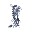
 Structure visualization
 Downloads & links
 Other downloads
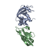

 PDBj
PDBj


 Assembly


 Mass: 74.922 Da / Num. of mol.: 2 / Source method: obtained synthetically / Formula: As
 Num. of mol.: 1 / Source method: obtained synthetically
 Mass: 18.015 Da / Num. of mol.: 11 / Source method: isolated from a natural source / Formula: H2O
 Sample preparation
 / Beamline: 19-ID / Wavelength: 0.97942 Å
 Processing