 Movie
Movie Controller

[English] 日本語
 Yorodumi
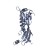
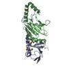
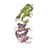
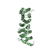
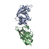
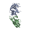
- PDB-5i7j: Crystal Structure of Human SPLUNC1 Disulfide Mutant M3 (I76C, V214C) -
+ Open data

- Basic information
| Entry | Database: PDB / ID: 5i7j | ||||||
|---|---|---|---|---|---|---|---|
| Title | Crystal Structure of Human SPLUNC1 Disulfide Mutant M3 (I76C, V214C) | ||||||
 Components Components | BPI fold-containing family A member 1 | ||||||
 Keywords Keywords | ANTIMICROBIAL PROTEIN / Innate pulmonary defense protein | ||||||
| Function / homology |  Function and homology information Function and homology informationimmune response in nasopharyngeal-associated lymphoid tissue / negative regulation of single-species biofilm formation in or on host organism / multicellular organismal-level water homeostasis / regulation of sodium ion transmembrane transport / surfactant homeostasis / Antimicrobial peptides / antimicrobial humoral immune response mediated by antimicrobial peptide / antibacterial humoral response / defense response to virus / innate immune response ...immune response in nasopharyngeal-associated lymphoid tissue / negative regulation of single-species biofilm formation in or on host organism / multicellular organismal-level water homeostasis / regulation of sodium ion transmembrane transport / surfactant homeostasis / Antimicrobial peptides / antimicrobial humoral immune response mediated by antimicrobial peptide / antibacterial humoral response / defense response to virus / innate immune response / lipid binding / : / extracellular region Similarity search - Function | ||||||
| Biological species |  Homo sapiens (human) Homo sapiens (human) | ||||||
| Method |  X-RAY DIFFRACTION / SYNCHROTRON / MOLECULAR REPLACEMENT / Resolution: 2.544 Å X-RAY DIFFRACTION / SYNCHROTRON / MOLECULAR REPLACEMENT / Resolution: 2.544 Å | ||||||
 Authors Authors | Walton, W.G. / Redinbo, M.R. | ||||||
| Funding support |  United States, 1items United States, 1items
| ||||||
 Citation Citation | Journal: Biochemistry / Year: 2016 Title: Structural Features Essential to the Antimicrobial Functions of Human SPLUNC1. Authors: Walton, W.G. / Ahmad, S. / Little, M.S. / Kim, C.S. / Tyrrell, J. / Lin, Q. / Di, Y.P. / Tarran, R. / Redinbo, M.R. | ||||||
| History |
|
- Structure visualization
| Structure viewer | Molecule: MolmilJmol/JSmol |
|---|
- Downloads & links
-Download
| PDBx/mmCIF format | 5i7j.cif.gz | 87.7 KB | Display | PDBx/mmCIF format |
|---|---|---|---|---|
| PDB format | pdb5i7j.ent.gz | 65.1 KB | Display | PDB format |
| PDBx/mmJSON format | 5i7j.json.gz | Tree view | PDBx/mmJSON format | |
| Others |  Other downloads Other downloads |
-Validation report
| Arichive directory | https://data.pdbj.org/pub/pdb/validation_reports/i7/5i7jftp://data.pdbj.org/pub/pdb/validation_reports/i7/5i7j | HTTPS FTP |
|---|
-Related structure data
| Related structure data |  5i7kC 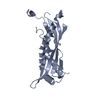 5i7lC 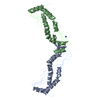 4kghS C: citing same article ( S: Starting model for refinement |
|---|---|
| Similar structure data |


-Links
 PDBj
PDBj

- Assembly
| Deposited unit | 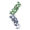
| ||||||||
|---|---|---|---|---|---|---|---|---|---|
| 1 | 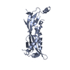
| ||||||||
| 2 | 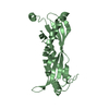
| ||||||||
| Unit cell |
|
-Components
| #1: Protein | Mass: 24953.320 Da / Num. of mol.: 2 / Mutation: I76C, V214C Source method: isolated from a genetically manipulated source Source: (gene. exp.) Homo sapiens (human)Gene: BPIFA1, LUNX, NASG, PLUNC, SPLUNC1, SPURT, UNQ787/PRO1606 Production host:  #2: Water | ChemComp-HOH / |  Mass: 18.015 Da / Num. of mol.: 13 / Source method: isolated from a natural source / Formula: H2O Mass: 18.015 Da / Num. of mol.: 13 / Source method: isolated from a natural source / Formula: H2OHas protein modification | Y | |
|---|
-Experimental details
-Experiment
| Experiment | Method: X-RAY DIFFRACTION / Number of used crystals: 1 |
|---|
- Sample preparation
| Crystal | Density Matthews: 2.9 Å3/Da / Density % sol: 57.62 % |
|---|---|
| Crystal grow | Temperature: 310 K / Method: vapor diffusion, hanging drop / pH: 8.5 / Details: 6M Ammonium Nitrate, 0.1M Tris-HCL, pH 8.5 |
-Data collection
| Diffraction | Mean temperature: 100 K |
|---|---|
| Diffraction source | Source: SYNCHROTRON / Site: APS / Beamline: 23-BM-B / Wavelength: 1.0332 Å |
| Detector | Type: MARMOSAIC 300 mm CCD / Detector: CCD / Date: Apr 23, 2015 |
| Radiation | Protocol: SINGLE WAVELENGTH / Monochromatic (M) / Laue (L): M / Scattering type: x-ray |
| Radiation wavelength | Wavelength: 1.0332 Å / Relative weight: 1 |
| Reflection | Resolution: 2.544→28.283 Å / Num. obs: 19395 / % possible obs: 98.74 % / Redundancy: 3.5 % / Biso Wilson estimate: 58.95 Å2 / CC1/2: 0.998 / Rmerge(I) obs: 0.05465 / Net I/σ(I): 14.54 |
| Reflection shell | Resolution: 2.544→2.634 Å / Redundancy: 3.4 % / Rmerge(I) obs: 0.7058 / % possible all: 94.45 |
- Processing
| Software |
| ||||||||||||||||||||||||||||||||||||||||||||||||||||||||||||||||||||||||||||||||||||||||||||||||||
|---|---|---|---|---|---|---|---|---|---|---|---|---|---|---|---|---|---|---|---|---|---|---|---|---|---|---|---|---|---|---|---|---|---|---|---|---|---|---|---|---|---|---|---|---|---|---|---|---|---|---|---|---|---|---|---|---|---|---|---|---|---|---|---|---|---|---|---|---|---|---|---|---|---|---|---|---|---|---|---|---|---|---|---|---|---|---|---|---|---|---|---|---|---|---|---|---|---|---|---|
| Refinement | Method to determine structure: MOLECULAR REPLACEMENT Starting model: 4KGH Resolution: 2.544→28.283 Å / SU ML: 0.36 / Cross valid method: FREE R-VALUE / σ(F): 0 / Phase error: 32.38
| ||||||||||||||||||||||||||||||||||||||||||||||||||||||||||||||||||||||||||||||||||||||||||||||||||
| Solvent computation | Shrinkage radii: 0.9 Å / VDW probe radii: 1.11 Å | ||||||||||||||||||||||||||||||||||||||||||||||||||||||||||||||||||||||||||||||||||||||||||||||||||
| Refinement step | Cycle: LAST / Resolution: 2.544→28.283 Å
| ||||||||||||||||||||||||||||||||||||||||||||||||||||||||||||||||||||||||||||||||||||||||||||||||||
| Refine LS restraints |
| ||||||||||||||||||||||||||||||||||||||||||||||||||||||||||||||||||||||||||||||||||||||||||||||||||
| LS refinement shell |
|
