 Movie
Movie Controller


+ Open data

- Basic information
| Entry | Database: PDB / ID: 2y9m | ||||||
|---|---|---|---|---|---|---|---|
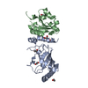
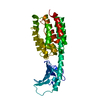
| Title | Pex4p-Pex22p structure | ||||||
 Components Components |
| ||||||
 Keywords Keywords | LIGASE/TRANSPORT PROTEIN / LIGASE-TRANSPORT PROTEIN COMPLEX / UBIQUITIN CONJUGATING ENZYME / E2 COMPLEX / PEROXISOMAL PROTEIN / ALPHA-BETA-ALPHA SANDWICH FOLD / E2 CO-ACTIVATOR | ||||||
| Function / homology |  Function and homology information Function and homology informationprotein import into peroxisome matrix, receptor recycling / peroxisome organization / ubiquitin-protein transferase activator activity / positive regulation of protein autoubiquitination / peroxisomal membrane / E2 ubiquitin-conjugating enzyme / ubiquitin conjugating enzyme activity / protein monoubiquitination / protein autoubiquitination / positive regulation of protein ubiquitination ...protein import into peroxisome matrix, receptor recycling / peroxisome organization / ubiquitin-protein transferase activator activity / positive regulation of protein autoubiquitination / peroxisomal membrane / E2 ubiquitin-conjugating enzyme / ubiquitin conjugating enzyme activity / protein monoubiquitination / protein autoubiquitination / positive regulation of protein ubiquitination / protein polyubiquitination / ubiquitin-protein transferase activity / peroxisome / protein ubiquitination / ATP binding / nucleus / cytoplasm Similarity search - Function | ||||||
| Biological species |  | ||||||
| Method |  X-RAY DIFFRACTION / SYNCHROTRON / MAD / Resolution: 2.6 Å X-RAY DIFFRACTION / SYNCHROTRON / MAD / Resolution: 2.6 Å | ||||||
 Authors Authors | Williams, C. / van den Berg, M. / Panjikar, S. / Distel, B. / Wilmanns, M. | ||||||
 Citation Citation | Journal: Embo J. / Year: 2011 Title: Insights Into Ubiquitin-Conjugating Enzyme/ Co-Activator Interactions from the Structure of the Pex4P:Pex22P Complex. Authors: Williams, C. / van den Berg, M. / Panjikar, S. / Stanley, W.A. / Distel, B. / Wilmanns, M. | ||||||
| History |
|
- Structure visualization
| Structure viewer | Molecule: MolmilJmol/JSmol |
|---|
- Downloads & links
-Download
| PDBx/mmCIF format | 2y9m.cif.gz | 126.7 KB | Display | PDBx/mmCIF format |
|---|---|---|---|---|
| PDB format | pdb2y9m.ent.gz | 99.4 KB | Display | PDB format |
| PDBx/mmJSON format | 2y9m.json.gz | Tree view | PDBx/mmJSON format | |
| Others |  Other downloads Other downloads |
-Validation report
| Arichive directory | https://data.pdbj.org/pub/pdb/validation_reports/y9/2y9mftp://data.pdbj.org/pub/pdb/validation_reports/y9/2y9m | HTTPS FTP |
|---|
-Related structure data
| Related structure data | |
|---|---|
| Similar structure data |


-Links
 PDBj
PDBj

- Assembly
| Deposited unit | 
| ||||||||
|---|---|---|---|---|---|---|---|---|---|
| 1 |
| ||||||||
| Unit cell |
|
-Components
| #1: Protein | Mass: 19573.463 Da / Num. of mol.: 1 / Fragment: UBC DOMAIN, RESIDUES 15-183 / Mutation: YES Source method: isolated from a genetically manipulated source Details: DISULPHIDE BOND BETWEEN RESIDUES 105 AND 146 Source: (gene. exp.) Plasmid: PCW187 / Production host:  | ||||||||
|---|---|---|---|---|---|---|---|---|---|
| #2: Protein | Mass: 14439.574 Da / Num. of mol.: 1 / Fragment: SOLUBLE DOMAIN, RESIDUES 54-180 Source method: isolated from a genetically manipulated source Source: (gene. exp.) Plasmid: PCW218 / Production host: | ||||||||
| #3: Chemical | ChemComp-EDO /   Mass: 62.068 Da / Num. of mol.: 4 / Source method: obtained synthetically / Formula: C2H6O2 Mass: 62.068 Da / Num. of mol.: 4 / Source method: obtained synthetically / Formula: C2H6O2#4: Water | ChemComp-HOH / |  Mass: 18.015 Da / Num. of mol.: 9 / Source method: isolated from a natural source / Formula: H2O Mass: 18.015 Da / Num. of mol.: 9 / Source method: isolated from a natural source / Formula: H2OCompound details | ENGINEERED | Has protein modification | Y | Sequence details | RESIDUE 15 IS MUTATED FROM SERINE TO ALANINE TO AID CLONING. THE N-TERMINAL GLYCINE IS LEFT OVER ...RESIDUE 15 IS MUTATED FROM SERINE TO ALANINE TO AID CLONING. THE N-TERMINAL GLYCINE IS LEFT OVER FROM THE TAG THE N-TERMINAL GLYCINE AND ALANINE ARE LEFT OVER FROM THE TAG | |
-Experimental details
-Experiment
| Experiment | Method: X-RAY DIFFRACTION / Number of used crystals: 1 |
|---|
- Sample preparation
| Crystal | Density Matthews: 2.76 Å3/Da / Density % sol: 55.46 % Description: A 4 WAVELENGTH MAD DATA SET WAS COLLECTED AND THE MODEL FROM THIS DATASET WAS USED AS MR MODEL FOR THE NATIVE DATASET |
|---|---|
| Crystal grow | pH: 7 Details: 8.0 % (V/V) ETHYLENE GLYCOL, 14.0 % (W/V) POLYETHYLENE GLYCOL, 0.1 M HEPES PH 7.0 |
-Data collection
| Diffraction | Mean temperature: 100 K |
|---|---|
| Diffraction source | Source: SYNCHROTRON / Site: ESRF  / Beamline: ID14-1 / Wavelength: 0.9334 / Beamline: ID14-1 / Wavelength: 0.9334 |
| Detector | Type: ADSC QUANTUM 210 / Detector: CCD / Date: Nov 20, 2008 / Details: DIAMOND 111 |
| Radiation | Monochromator: DIAMOND 111 / Protocol: MAD / Monochromatic (M) / Laue (L): M / Scattering type: x-ray |
| Radiation wavelength | Wavelength: 0.9334 Å / Relative weight: 1 |
| Reflection | Resolution: 2.6→34.7 Å / Num. obs: 11039 / % possible obs: 99.4 % / Observed criterion σ(I): 3 / Redundancy: 7.5 % / Biso Wilson estimate: 52 Å2 / Rmerge(I) obs: 0.06 / Net I/σ(I): 26.7 |
| Reflection shell | Resolution: 2.6→2.74 Å / Redundancy: 7.6 % / Rmerge(I) obs: 0.47 / Mean I/σ(I) obs: 4.8 / % possible all: 99.1 |
- Processing
| Software |
| ||||||||||||||||||||||||||||||||||||||||||||||||||||||||||||||||||||||||||||||||||||||||||||||||||||||||||||||||||||||||||||||||||||||||||||||||||||||||||||||||||||||||||||||||||||||
|---|---|---|---|---|---|---|---|---|---|---|---|---|---|---|---|---|---|---|---|---|---|---|---|---|---|---|---|---|---|---|---|---|---|---|---|---|---|---|---|---|---|---|---|---|---|---|---|---|---|---|---|---|---|---|---|---|---|---|---|---|---|---|---|---|---|---|---|---|---|---|---|---|---|---|---|---|---|---|---|---|---|---|---|---|---|---|---|---|---|---|---|---|---|---|---|---|---|---|---|---|---|---|---|---|---|---|---|---|---|---|---|---|---|---|---|---|---|---|---|---|---|---|---|---|---|---|---|---|---|---|---|---|---|---|---|---|---|---|---|---|---|---|---|---|---|---|---|---|---|---|---|---|---|---|---|---|---|---|---|---|---|---|---|---|---|---|---|---|---|---|---|---|---|---|---|---|---|---|---|---|---|---|---|
| Refinement | Method to determine structure: MAD Starting model: NONE Resolution: 2.6→20 Å / Cor.coef. Fo:Fc: 0.941 / Cor.coef. Fo:Fc free: 0.897 / SU B: 22.417 / SU ML: 0.228 / Cross valid method: THROUGHOUT / ESU R: 0.38 / ESU R Free: 0.321 / Stereochemistry target values: MAXIMUM LIKELIHOOD / Details: HYDROGENS HAVE BEEN ADDED IN THE RIDING POSITIONS.
| ||||||||||||||||||||||||||||||||||||||||||||||||||||||||||||||||||||||||||||||||||||||||||||||||||||||||||||||||||||||||||||||||||||||||||||||||||||||||||||||||||||||||||||||||||||||
| Solvent computation | Ion probe radii: 0.8 Å / Shrinkage radii: 0.8 Å / VDW probe radii: 1.2 Å / Solvent model: MASK | ||||||||||||||||||||||||||||||||||||||||||||||||||||||||||||||||||||||||||||||||||||||||||||||||||||||||||||||||||||||||||||||||||||||||||||||||||||||||||||||||||||||||||||||||||||||
| Displacement parameters | Biso mean: 46.852 Å2
| ||||||||||||||||||||||||||||||||||||||||||||||||||||||||||||||||||||||||||||||||||||||||||||||||||||||||||||||||||||||||||||||||||||||||||||||||||||||||||||||||||||||||||||||||||||||
| Refinement step | Cycle: LAST / Resolution: 2.6→20 Å
| ||||||||||||||||||||||||||||||||||||||||||||||||||||||||||||||||||||||||||||||||||||||||||||||||||||||||||||||||||||||||||||||||||||||||||||||||||||||||||||||||||||||||||||||||||||||
| Refine LS restraints |
|