 Movie
Movie Controller


+ Open data

- Basic information
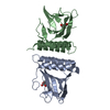
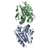
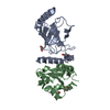
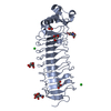
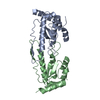
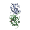
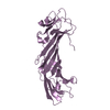
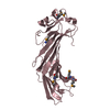
| Entry | Database: PDB / ID: 1a87 | ||||||
|---|---|---|---|---|---|---|---|
| Title | COLICIN N | ||||||
 Components Components | COLICIN N | ||||||
 Keywords Keywords | BACTERIOCIN / TOXIN / PORE-FORMING ACTIVITY | ||||||
| Function / homology |  Function and homology information Function and homology informationpore-forming activity / killing of cells of another organism / defense response to Gram-negative bacterium / transmembrane transporter binding / defense response to bacterium / plasma membrane Similarity search - Function | ||||||
| Biological species |  | ||||||
| Method |  X-RAY DIFFRACTION / SYNCHROTRON / MIR / Resolution: 3.1 Å X-RAY DIFFRACTION / SYNCHROTRON / MIR / Resolution: 3.1 Å | ||||||
 Authors Authors | Vetter, I.R. / Parker, M.W. / Tucker, A.D. / Lakey, J.H. / Pattus, F. / Tsernoglou, D. | ||||||
 Citation Citation | Journal: Structure / Year: 1998 Title: Crystal structure of a colicin N fragment suggests a model for toxicity. Authors: Vetter, I.R. / Parker, M.W. / Tucker, A.D. / Lakey, J.H. / Pattus, F. / Tsernoglou, D. #1: Journal: Protein Toxin Structure / Year: 1996Title: Insights Into Membrane Insertion Based on Studies of Colicins Authors: Vetter, I.R. / Parker, M.W. / Pattus, F. / Tsernoglou, D. #2: Journal: Eur.J.Biochem. / Year: 1993Title: Characterization of the Receptor and Translocator Domains of Colicin N Authors: El Kouhen, R. / Fierobe, H.P. / Scianimanico, S. / Steiert, M. / Pattus, F. / Pages, J.M. #3: Journal: Eur.Biophys.J. / Year: 1990Title: Colicin N Forms Voltage-and Ph-Dependent Channels in Planar Lipid Bilayer Membranes Authors: Wilmsen, H.U. / Pugsley, A.P. / Pattus, F. #4: Journal: Mol.Microbiol. / Year: 1990Title: Involvement of Ompf During Reception and Translocation Steps of Colicin N Entry Authors: Bourdineaud, J.P. / Fierobe, H.P. / Lazdunski, C. / Pages, J.M. #5: Journal: Mol.Microbiol. / Year: 1987Title: Nucleotide Sequencing of the Structural Gene for Colicin N Reveals Homology between the Catalytic, C-Terminal Domains of Colicins a and N Authors: Pugsley, A.P. | ||||||
| History |
|
- Structure visualization
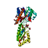
| Structure viewer | Molecule: MolmilJmol/JSmol |
|---|
- Downloads & links
-Download
| PDBx/mmCIF format | 1a87.cif.gz | 70.5 KB | Display | PDBx/mmCIF format |
|---|---|---|---|---|
| PDB format | pdb1a87.ent.gz | 53.3 KB | Display | PDB format |
| PDBx/mmJSON format | 1a87.json.gz | Tree view | PDBx/mmJSON format | |
| Others |  Other downloads Other downloads |
-Validation report
| Arichive directory | https://data.pdbj.org/pub/pdb/validation_reports/a8/1a87ftp://data.pdbj.org/pub/pdb/validation_reports/a8/1a87 | HTTPS FTP |
|---|
-Related structure data
| Similar structure data |
|---|

-Links
 PDBj
PDBj- Assembly
| Deposited unit | 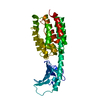
| ||||||||
|---|---|---|---|---|---|---|---|---|---|
| 1 |
| ||||||||
| Unit cell |
|
-Components
| #1: Protein | Mass: 35146.270 Da / Num. of mol.: 1 / Fragment: RECEPTOR-BINDING AND PORE-FORMING DOMAIN Source method: isolated from a genetically manipulated source Source: (gene. exp.) |
|---|---|
| #2: Water | ChemComp-HOH /  Mass: 18.015 Da / Num. of mol.: 55 / Source method: isolated from a natural source / Formula: H2O Mass: 18.015 Da / Num. of mol.: 55 / Source method: isolated from a natural source / Formula: H2O |
-Experimental details
-Experiment
| Experiment | Method: X-RAY DIFFRACTION / Number of used crystals: 1 |
|---|
- Sample preparation
| Crystal | Density Matthews: 4.17 Å3/Da / Density % sol: 65 % | ||||||||||||||||||||
|---|---|---|---|---|---|---|---|---|---|---|---|---|---|---|---|---|---|---|---|---|---|
| Crystal grow | *PLUS Method: vapor diffusion, hanging drop / PH range low: 7 / PH range high: 5.6 | ||||||||||||||||||||
| Components of the solutions | *PLUS
|
-Data collection
| Diffraction | Mean temperature: 283 K |
|---|---|
| Diffraction source | Source: SYNCHROTRON / Site: EMBL/DESY, HAMBURG  / Beamline: X31 / Wavelength: 1.009 / Beamline: X31 / Wavelength: 1.009 |
| Detector | Type: MARRESEARCH / Detector: IMAGE PLATE / Date: Jun 1, 1990 |
| Radiation | Monochromatic (M) / Laue (L): M / Scattering type: x-ray |
| Radiation wavelength | Wavelength: 1.009 Å / Relative weight: 1 |
| Reflection | Resolution: 3.1→22.7 Å / Num. obs: 10644 / % possible obs: 93.5 % / Biso Wilson estimate: 57.5 Å2 / Rsym value: 0.083 / Net I/σ(I): 33.07 |
| Reflection shell | Resolution: 3.1→3.2 Å / Mean I/σ(I) obs: 6.3 / % possible all: 100 |
| Reflection | *PLUS Rmerge(I) obs: 0.083 |
| Reflection shell | *PLUS % possible obs: 100 % |
- Processing
| Software |
| ||||||||||||||||||||||||||||||||||||||||
|---|---|---|---|---|---|---|---|---|---|---|---|---|---|---|---|---|---|---|---|---|---|---|---|---|---|---|---|---|---|---|---|---|---|---|---|---|---|---|---|---|---|
| Refinement | Method to determine structure: MIR / Resolution: 3.1→23 Å / Isotropic thermal model: TNT BCORREL / Cross valid method: POSTERIORI / σ(F): 0 / Stereochemistry target values: TNT PROTGEO
| ||||||||||||||||||||||||||||||||||||||||
| Solvent computation | Bsol: 300 Å2 / ksol: 0.8 e/Å3 | ||||||||||||||||||||||||||||||||||||||||
| Refinement step | Cycle: LAST / Resolution: 3.1→23 Å
| ||||||||||||||||||||||||||||||||||||||||
| Refine LS restraints |
| ||||||||||||||||||||||||||||||||||||||||
| Software | *PLUS Name: TNT / Version: 5EB / Classification: refinement | ||||||||||||||||||||||||||||||||||||||||
| Refinement | *PLUS Rfactor obs: 0.173 | ||||||||||||||||||||||||||||||||||||||||
| Solvent computation | *PLUS | ||||||||||||||||||||||||||||||||||||||||
| Displacement parameters | *PLUS | ||||||||||||||||||||||||||||||||||||||||
| Refine LS restraints | *PLUS
|
