 Movie
Movie Controller

[English] 日本語
 Yorodumi
- PDB-5y3t: Crystal structure of hetero-trimeric core of LUBAC: HOIP double-U... -
+ Open data

- Basic information
| Entry | Database: PDB / ID: 5y3t | |||||||||
|---|---|---|---|---|---|---|---|---|---|---|
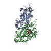
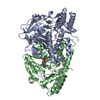
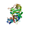
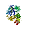
| Title | Crystal structure of hetero-trimeric core of LUBAC: HOIP double-UBA complexed with HOIL-1L UBL and SHARPIN UBL | |||||||||
 Components Components |
| |||||||||
 Keywords Keywords | LIGASE / HOIP / HOIL-1L / SHARPIN / Linear-Ubiquitin Assembly Complex / NF-KB Activation / LUBAC Tethering Motif / LUBAC Stabilization | |||||||||
| Function / homology |  Function and homology information Function and homology informationTNFR1-induced proapoptotic signaling / apoptotic nuclear changes / regulation of CD40 signaling pathway / protein linear polyubiquitination / LUBAC complex / TNFR1-induced NF-kappa-B signaling pathway / linear polyubiquitin binding / Regulation of TNFR1 signaling / RBR-type E3 ubiquitin transferase / CD40 signaling pathway ...TNFR1-induced proapoptotic signaling / apoptotic nuclear changes / regulation of CD40 signaling pathway / protein linear polyubiquitination / LUBAC complex / TNFR1-induced NF-kappa-B signaling pathway / linear polyubiquitin binding / Regulation of TNFR1 signaling / RBR-type E3 ubiquitin transferase / CD40 signaling pathway / positive regulation of xenophagy / CD40 receptor complex / negative regulation of necroptotic process / regulation of tumor necrosis factor-mediated signaling pathway / Antigen processing: Ubiquitination & Proteasome degradation / ubiquitin ligase activator activity / positive regulation of extrinsic apoptotic signaling pathway / K48-linked polyubiquitin modification-dependent protein binding / K63-linked polyubiquitin modification-dependent protein binding / keratinization / epidermis development / polyubiquitin modification-dependent protein binding / protein kinase C binding / tumor necrosis factor-mediated signaling pathway / : / ubiquitin binding / mitochondrion organization / positive regulation of non-canonical NF-kappaB signal transduction / negative regulation of inflammatory response / cytoplasmic side of plasma membrane / protein polyubiquitination / ubiquitin-protein transferase activity / ubiquitin protein ligase activity / T cell receptor signaling pathway / double-stranded DNA binding / proteasome-mediated ubiquitin-dependent protein catabolic process / protein-macromolecule adaptor activity / positive regulation of canonical NF-kappaB signal transduction / defense response to bacterium / protein ubiquitination / positive regulation of apoptotic process / ubiquitin protein ligase binding / synapse / dendrite / protein-containing complex binding / positive regulation of transcription by RNA polymerase II / zinc ion binding / metal ion binding / identical protein binding / cytosol Similarity search - Function | |||||||||
| Biological species |  | |||||||||
| Method |  X-RAY DIFFRACTION / SYNCHROTRON / MOLECULAR REPLACEMENT / Resolution: 2.4 Å X-RAY DIFFRACTION / SYNCHROTRON / MOLECULAR REPLACEMENT / Resolution: 2.4 Å | |||||||||
 Authors Authors | Tokunaga, A. / Fujita, H. / Ariyoshi, M. / Ohki, I. / Tochio, H. / Iwai, K. / Shirakawa, M. | |||||||||
| Funding support |  Japan, 2items Japan, 2items
| |||||||||
 Citation Citation | Journal: Cell Rep / Year: 2018 Title: Cooperative Domain Formation by Homologous Motifs in HOIL-1L and SHARPIN Plays A Crucial Role in LUBAC Stabilization. Authors: Fujita, H. / Tokunaga, A. / Shimizu, S. / Whiting, A.L. / Aguilar-Alonso, F. / Takagi, K. / Walinda, E. / Sasaki, Y. / Shimokawa, T. / Mizushima, T. / Ohki, I. / Ariyoshi, M. / Tochio, H. / ...Authors: Fujita, H. / Tokunaga, A. / Shimizu, S. / Whiting, A.L. / Aguilar-Alonso, F. / Takagi, K. / Walinda, E. / Sasaki, Y. / Shimokawa, T. / Mizushima, T. / Ohki, I. / Ariyoshi, M. / Tochio, H. / Bernal, F. / Shirakawa, M. / Iwai, K. | |||||||||
| History |
|
- Structure visualization
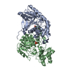
| Structure viewer | Molecule: MolmilJmol/JSmol |
|---|
- Downloads & links
-Download
| PDBx/mmCIF format | 5y3t.cif.gz | 99.1 KB | Display | PDBx/mmCIF format |
|---|---|---|---|---|
| PDB format | pdb5y3t.ent.gz | 73.7 KB | Display | PDB format |
| PDBx/mmJSON format | 5y3t.json.gz | Tree view | PDBx/mmJSON format | |
| Others |  Other downloads Other downloads |
-Validation report
| Arichive directory | https://data.pdbj.org/pub/pdb/validation_reports/y3/5y3tftp://data.pdbj.org/pub/pdb/validation_reports/y3/5y3t | HTTPS FTP |
|---|
-Related structure data
| Related structure data |  4dbgS S: Starting model for refinement |
|---|---|
| Similar structure data |


-Links
 PDBj
PDBj


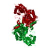
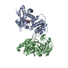
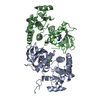
- Assembly
| Deposited unit | 
| ||||||||
|---|---|---|---|---|---|---|---|---|---|
| 1 |
| ||||||||
| Unit cell |
|
-Components
| #1: Protein | Mass: 15762.995 Da / Num. of mol.: 1 / Fragment: UNP residues 1-140 Source method: isolated from a genetically manipulated source Source: (gene. exp.)  References: UniProt: Q9WUB0, RING-type E3 ubiquitin transferase |
|---|---|
| #2: Protein | Mass: 17995.115 Da / Num. of mol.: 1 / Fragment: UNP residues 474-630 Source method: isolated from a genetically manipulated source Source: (gene. exp.) References: UniProt: Q924T7, RING-type E3 ubiquitin transferase |
| #3: Protein | Mass: 19921.514 Da / Num. of mol.: 1 / Fragment: UNP residues 163-341 Source method: isolated from a genetically manipulated source Source: (gene. exp.) |
| #4: Chemical | ChemComp-GOL /   Mass: 92.094 Da / Num. of mol.: 1 / Source method: obtained synthetically / Formula: C3H8O3 Mass: 92.094 Da / Num. of mol.: 1 / Source method: obtained synthetically / Formula: C3H8O3 |
| #5: Water | ChemComp-HOH /  Mass: 18.015 Da / Num. of mol.: 21 / Source method: isolated from a natural source / Formula: H2O Mass: 18.015 Da / Num. of mol.: 21 / Source method: isolated from a natural source / Formula: H2O |
-Experimental details
-Experiment
| Experiment | Method: X-RAY DIFFRACTION / Number of used crystals: 1 |
|---|
- Sample preparation
| Crystal | Density Matthews: 2.33 Å3/Da / Density % sol: 47.14 % |
|---|---|
| Crystal grow | Temperature: 293 K / Method: vapor diffusion, sitting drop / pH: 6.5 / Details: Magnesium sulfate, MES |
-Data collection
| Diffraction | Mean temperature: 95 K |
|---|---|
| Diffraction source | Source: SYNCHROTRON / Site: Photon Factory / Beamline: BL-17A / Wavelength: 0.98 Å |
| Detector | Type: ADSC QUANTUM 270 / Detector: CCD / Date: Mar 7, 2017 |
| Radiation | Monochromator: Si(111) crystals / Protocol: SINGLE WAVELENGTH / Monochromatic (M) / Laue (L): M / Scattering type: x-ray |
| Radiation wavelength | Wavelength: 0.98 Å / Relative weight: 1 |
| Reflection | Resolution: 2.4→50 Å / Num. obs: 19275 / % possible obs: 99.3 % / Redundancy: 4.5 % / Rmerge(I) obs: 0.092 / Net I/σ(I): 14.2 |
| Reflection shell | Resolution: 2.4→2.44 Å / Redundancy: 4.2 % / Rmerge(I) obs: 0.52 / Mean I/σ(I) obs: 1.9 / % possible all: 95.5 |
- Processing
| Software |
| ||||||||||||||||||||||||||||||||||||||||||||||||||||||||||||||||||||||||||||||||||||||||||||||||||||||||||||||||||||||||||||||||||||||||||||||||||||||||||||||||||||||||||||||||||||||
|---|---|---|---|---|---|---|---|---|---|---|---|---|---|---|---|---|---|---|---|---|---|---|---|---|---|---|---|---|---|---|---|---|---|---|---|---|---|---|---|---|---|---|---|---|---|---|---|---|---|---|---|---|---|---|---|---|---|---|---|---|---|---|---|---|---|---|---|---|---|---|---|---|---|---|---|---|---|---|---|---|---|---|---|---|---|---|---|---|---|---|---|---|---|---|---|---|---|---|---|---|---|---|---|---|---|---|---|---|---|---|---|---|---|---|---|---|---|---|---|---|---|---|---|---|---|---|---|---|---|---|---|---|---|---|---|---|---|---|---|---|---|---|---|---|---|---|---|---|---|---|---|---|---|---|---|---|---|---|---|---|---|---|---|---|---|---|---|---|---|---|---|---|---|---|---|---|---|---|---|---|---|---|---|
| Refinement | Method to determine structure: MOLECULAR REPLACEMENT Starting model: 4DBG Resolution: 2.4→48.19 Å / Cor.coef. Fo:Fc: 0.964 / Cor.coef. Fo:Fc free: 0.942 / SU B: 9.857 / SU ML: 0.22 / Cross valid method: THROUGHOUT / ESU R: 0.396 / ESU R Free: 0.256 / Stereochemistry target values: MAXIMUM LIKELIHOOD / Details: HYDROGENS HAVE BEEN ADDED IN THE RIDING POSITIONS
| ||||||||||||||||||||||||||||||||||||||||||||||||||||||||||||||||||||||||||||||||||||||||||||||||||||||||||||||||||||||||||||||||||||||||||||||||||||||||||||||||||||||||||||||||||||||
| Solvent computation | Ion probe radii: 0.8 Å / Shrinkage radii: 0.8 Å / VDW probe radii: 1.2 Å / Solvent model: MASK | ||||||||||||||||||||||||||||||||||||||||||||||||||||||||||||||||||||||||||||||||||||||||||||||||||||||||||||||||||||||||||||||||||||||||||||||||||||||||||||||||||||||||||||||||||||||
| Displacement parameters | Biso mean: 65.224 Å2
| ||||||||||||||||||||||||||||||||||||||||||||||||||||||||||||||||||||||||||||||||||||||||||||||||||||||||||||||||||||||||||||||||||||||||||||||||||||||||||||||||||||||||||||||||||||||
| Refinement step | Cycle: 1 / Resolution: 2.4→48.19 Å
| ||||||||||||||||||||||||||||||||||||||||||||||||||||||||||||||||||||||||||||||||||||||||||||||||||||||||||||||||||||||||||||||||||||||||||||||||||||||||||||||||||||||||||||||||||||||
| Refine LS restraints |
|