 Movie
Movie Controller

[English] 日本語
 Yorodumi
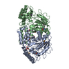
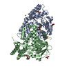
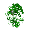
- PDB-1fg7: CRYSTAL STRUCTURE OF L-HISTIDINOL PHOSPHATE AMINOTRANSFERASE WITH... -
+ Open data

- Basic information
| Entry | Database: PDB / ID: 1fg7 | ||||||
|---|---|---|---|---|---|---|---|
| Title | CRYSTAL STRUCTURE OF L-HISTIDINOL PHOSPHATE AMINOTRANSFERASE WITH PYRIDOXAL-5'-PHOSPHATE | ||||||
 Components Components | HISTIDINOL PHOSPHATE AMINOTRANSFERASE | ||||||
 Keywords Keywords | TRANSFERASE / Aminotransferase / HisC / Histidine Biosynthesis / Pyridoxal Phosphate / Montreal-Kingston Bacterial Structural Genomics Initiative / BSGI / Structural Genomics | ||||||
| Function / homology |  Function and homology information Function and homology informationhistidinol-phosphate transaminase / L-histidinol-phosphate:2-oxoglutarate transaminase activity / L-histidine biosynthetic process / pyridoxal phosphate binding / identical protein binding / cytosol Similarity search - Function | ||||||
| Biological species |  | ||||||
| Method |  X-RAY DIFFRACTION / SYNCHROTRON / Resolution: 1.5 Å X-RAY DIFFRACTION / SYNCHROTRON / Resolution: 1.5 Å | ||||||
 Authors Authors | Sivaraman, J. / Cygler, M. / Montreal-Kingston Bacterial Structural Genomics Initiative (BSGI) | ||||||
 Citation Citation | Journal: J.Mol.Biol. / Year: 2001 Title: Crystal structure of histidinol phosphate aminotransferase (HisC) from Escherichia coli, and its covalent complex with pyridoxal-5'-phosphate and l-histidinol phosphate. Authors: Sivaraman, J. / Li, Y. / Larocque, R. / Schrag, J.D. / Cygler, M. / Matte, A. | ||||||
| History |
|
- Structure visualization
| Structure viewer | Molecule: MolmilJmol/JSmol |
|---|
- Downloads & links
-Download
| PDBx/mmCIF format | 1fg7.cif.gz | 85 KB | Display | PDBx/mmCIF format |
|---|---|---|---|---|
| PDB format | pdb1fg7.ent.gz | 63.7 KB | Display | PDB format |
| PDBx/mmJSON format | 1fg7.json.gz | Tree view | PDBx/mmJSON format | |
| Others |  Other downloads Other downloads |
-Validation report
| Arichive directory | https://data.pdbj.org/pub/pdb/validation_reports/fg/1fg7ftp://data.pdbj.org/pub/pdb/validation_reports/fg/1fg7 | HTTPS FTP |
|---|
-Related structure data


-Links
 PDBj
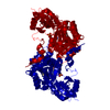
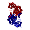
PDBj- Assembly
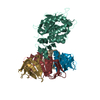
| Deposited unit | 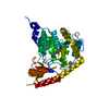
| ||||||||
|---|---|---|---|---|---|---|---|---|---|
| 1 | 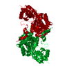
| ||||||||
| Unit cell |
|
-Components
| #1: Protein | Mass: 39581.562 Da / Num. of mol.: 1 Source method: isolated from a genetically manipulated source Source: (gene. exp.) References: UniProt: P06986, histidinol-phosphate transaminase |
|---|---|
| #2: Chemical | ChemComp-PMP /   Mass: 248.173 Da / Num. of mol.: 1 / Source method: obtained synthetically / Formula: C8H13N2O5P Mass: 248.173 Da / Num. of mol.: 1 / Source method: obtained synthetically / Formula: C8H13N2O5P |
| #3: Water | ChemComp-HOH /  Mass: 18.015 Da / Num. of mol.: 224 / Source method: isolated from a natural source / Formula: H2O Mass: 18.015 Da / Num. of mol.: 224 / Source method: isolated from a natural source / Formula: H2O |
| Has protein modification | Y |
-Experimental details
-Experiment
| Experiment | Method: X-RAY DIFFRACTION / Number of used crystals: 1 |
|---|
- Sample preparation
| Crystal | Density Matthews: 2.29 Å3/Da / Density % sol: 46.35 % | ||||||||||||||||||||||||||||||||||||||||||||||||||||||
|---|---|---|---|---|---|---|---|---|---|---|---|---|---|---|---|---|---|---|---|---|---|---|---|---|---|---|---|---|---|---|---|---|---|---|---|---|---|---|---|---|---|---|---|---|---|---|---|---|---|---|---|---|---|---|---|
| Crystal grow | Temperature: 292 K / Method: vapor diffusion, hanging drop / pH: 8.5 Details: PEG 3350, Tris-Hcl, MgCl2, Glycerol, pH 8.5, VAPOR DIFFUSION, HANGING DROP, temperature 292K | ||||||||||||||||||||||||||||||||||||||||||||||||||||||
| Crystal grow | *PLUS pH: 7.5 | ||||||||||||||||||||||||||||||||||||||||||||||||||||||
| Components of the solutions | *PLUS
|
-Data collection
| Diffraction | Mean temperature: 100 K |
|---|---|
| Diffraction source | Source: SYNCHROTRON / Site: NSLS  / Beamline: X8C / Wavelength: 0.961123 / Beamline: X8C / Wavelength: 0.961123 |
| Detector | Type: ADSC QUANTUM / Detector: CCD / Date: Mar 27, 2000 |
| Radiation | Protocol: SINGLE WAVELENGTH / Monochromatic (M) / Laue (L): M / Scattering type: x-ray |
| Radiation wavelength | Wavelength: 0.961123 Å / Relative weight: 1 |
| Reflection | Resolution: 1.4→100 Å / Num. all: 504180 / Num. obs: 137314 / % possible obs: 99.1 % / Observed criterion σ(I): 0.3 / Redundancy: 4 % / Biso Wilson estimate: 12.8 Å2 / Rmerge(I) obs: 0.05 / Net I/σ(I): 12 |
| Reflection shell | Resolution: 1.4→1.45 Å / Redundancy: 4 % / Rmerge(I) obs: 0.251 / Num. unique all: 13543 / % possible all: 97.5 |
| Reflection | *PLUS Num. measured all: 504180 |
- Processing
| Software |
| ||||||||||||||||
|---|---|---|---|---|---|---|---|---|---|---|---|---|---|---|---|---|---|
| Refinement | Resolution: 1.5→45 Å / Cross valid method: THROUGHOUT / σ(F): 0 / σ(I): 0 / Stereochemistry target values: Engh & Huber
| ||||||||||||||||
| Refinement step | Cycle: LAST / Resolution: 1.5→45 Å
| ||||||||||||||||
| Refine LS restraints |
| ||||||||||||||||
| Software | *PLUS Name: CNS / Classification: refinement | ||||||||||||||||
| Refinement | *PLUS Highest resolution: 1.5 Å / Lowest resolution: 45 Å / σ(F): 0 / Rfactor obs: 0.205 | ||||||||||||||||
| Solvent computation | *PLUS | ||||||||||||||||
| Displacement parameters | *PLUS | ||||||||||||||||
| Refine LS restraints | *PLUS
|
