 Movie
Movie Controller

[English] 日本語
 Yorodumi
- PDB-2c0r: CRYSTAL STRUCTURE OF PHOSPHOSERINE AMINOTRANSFERASE FROM BACILLUS... -
+ Open data

- Basic information
| Entry | Database: PDB / ID: 2c0r | ||||||
|---|---|---|---|---|---|---|---|
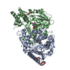
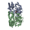
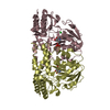
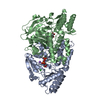
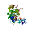
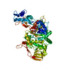
| Title | CRYSTAL STRUCTURE OF PHOSPHOSERINE AMINOTRANSFERASE FROM BACILLUS CIRCULANS VAR. ALKALOPHILUS AT pH 8.5 | ||||||
 Components Components | PHOSPHOSERINE AMINOTRANSFERASE | ||||||
 Keywords Keywords | TRANSFERASE / PHOSPHOSERINE AMINOTRANSFERASE / PYRIDOXAL-5'-PHOSPHATE / PYRIDINE SERINE BIOSYNTHESIS / AMINO-ACID BIOSYNTHESIS / AMINOTRANSFERASE / PYRIDOXAL PHOSPHATE / PYRIDOXINE BIOSYNTHESIS / SERINE BIOSYNTHESIS | ||||||
| Function / homology |  Function and homology information Function and homology informationphosphoserine transaminase / O-phospho-L-serine:2-oxoglutarate transaminase activity / L-serine biosynthetic process / pyridoxal phosphate binding / cytoplasm Similarity search - Function | ||||||
| Biological species |  BACILLUS CIRCULANS (bacteria) BACILLUS CIRCULANS (bacteria) | ||||||
| Method |  X-RAY DIFFRACTION / SYNCHROTRON / MOLECULAR REPLACEMENT / Resolution: 1.2 Å X-RAY DIFFRACTION / SYNCHROTRON / MOLECULAR REPLACEMENT / Resolution: 1.2 Å | ||||||
 Authors Authors | Kapetaniou, E.G. / Papageorgiou, A.C. | ||||||
 Citation Citation | Journal: Proteins: Struct., Funct., Bioinf. / Year: 2006 Title: Effect of Ph on the Structure and Stability of Bacillus Circulans Ssp. Alkalophilus Phosphoserine Aminotransferase: Thermodynamic and Crystallographic Studies. Authors: Kapetaniou, E.G. / Thanassoulas, A. / Dubnovitsky, A.P. / Nounesis, G. / Papageorgiou, A.C. | ||||||
| History |
|
- Structure visualization
| Structure viewer | Molecule: MolmilJmol/JSmol |
|---|
- Downloads & links
-Download
| PDBx/mmCIF format | 2c0r.cif.gz | 318.4 KB | Display | PDBx/mmCIF format |
|---|---|---|---|---|
| PDB format | pdb2c0r.ent.gz | 259.2 KB | Display | PDB format |
| PDBx/mmJSON format | 2c0r.json.gz | Tree view | PDBx/mmJSON format | |
| Others |  Other downloads Other downloads |
-Validation report
| Arichive directory | https://data.pdbj.org/pub/pdb/validation_reports/c0/2c0rftp://data.pdbj.org/pub/pdb/validation_reports/c0/2c0r | HTTPS FTP |
|---|
-Related structure data
| Related structure data | 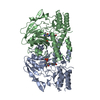 1bjnS S: Starting model for refinement |
|---|---|
| Similar structure data |


-Links
 PDBj
PDBj- Assembly
| Deposited unit | 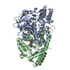
| ||||||||
|---|---|---|---|---|---|---|---|---|---|
| 1 |
| ||||||||
| Unit cell |
| ||||||||
| Noncrystallographic symmetry (NCS) | NCS oper: (Code: given Matrix: (-0.99995, 0.0063, 0.0079), Vector: |
-Components
| #1: Protein | Mass: 39966.090 Da / Num. of mol.: 2 / Mutation: YES Source method: isolated from a genetically manipulated source Source: (gene. exp.) BACILLUS CIRCULANS (bacteria) / Variant: ALKALOPHILUS / Production host: #2: Chemical |   Mass: 247.142 Da / Num. of mol.: 2 / Source method: obtained synthetically / Formula: C8H10NO6P Mass: 247.142 Da / Num. of mol.: 2 / Source method: obtained synthetically / Formula: C8H10NO6P#3: Water | ChemComp-HOH / |  Mass: 18.015 Da / Num. of mol.: 809 / Source method: isolated from a natural source / Formula: H2O Mass: 18.015 Da / Num. of mol.: 809 / Source method: isolated from a natural source / Formula: H2OCompound details | ENGINEERED | |
|---|
-Experimental details
-Experiment
| Experiment | Method: X-RAY DIFFRACTION / Number of used crystals: 1 |
|---|
- Sample preparation
| Crystal | Density Matthews: 2 Å3/Da / Density % sol: 38 % |
|---|---|
| Crystal grow | pH: 8.5 Details: 30% PEG 4000, 0.1 M TRIS-HCL BUFFER, PH 8.5, 5% GLYCEROL, 0.2 M SODIUM ACETATE |
-Data collection
| Diffraction | Mean temperature: 100 K |
|---|---|
| Diffraction source | Source: SYNCHROTRON / Site: EMBL/DESY, HAMBURG  / Beamline: X11 / Wavelength: 0.81 / Beamline: X11 / Wavelength: 0.81 |
| Detector | Type: MARRESEARCH / Detector: CCD / Date: Jul 4, 2003 |
| Radiation | Protocol: SINGLE WAVELENGTH / Monochromatic (M) / Laue (L): M / Scattering type: x-ray |
| Radiation wavelength | Wavelength: 0.81 Å / Relative weight: 1 |
| Reflection | Resolution: 1.2→15 Å / Num. obs: 171731 / % possible obs: 85.7 % / Observed criterion σ(I): 3 / Redundancy: 24.86 % / Rmerge(I) obs: 0.08 / Net I/σ(I): 18.83 |
| Reflection shell | Resolution: 1.2→1.24 Å / Rmerge(I) obs: 0.51 / Mean I/σ(I) obs: 2.4 / % possible all: 74.4 |
- Processing
| Software |
| |||||||||||||||||||||||||||||||||
|---|---|---|---|---|---|---|---|---|---|---|---|---|---|---|---|---|---|---|---|---|---|---|---|---|---|---|---|---|---|---|---|---|---|---|
| Refinement | Method to determine structure: MOLECULAR REPLACEMENT Starting model: PDB ENTRY 1BJN Resolution: 1.2→12 Å / Num. parameters: 57709 / Num. restraintsaints: 69730 / Cross valid method: FREE R-VALUE / σ(F): 0 / Stereochemistry target values: ENGH AND HUBER / Details: ANISOTROPIC REFINEMENT
| |||||||||||||||||||||||||||||||||
| Refine analyze | Num. disordered residues: 4 / Occupancy sum hydrogen: 0 / Occupancy sum non hydrogen: 6397 | |||||||||||||||||||||||||||||||||
| Refinement step | Cycle: LAST / Resolution: 1.2→12 Å
| |||||||||||||||||||||||||||||||||
| Refine LS restraints |
|
