 Movie
Movie Controller

[English] 日本語
 Yorodumi
- PDB-1bjn: STRUCTURE OF PHOSPHOSERINE AMINOTRANSFERASE FROM ESCHERICHIA COLI -
+ Open data

- Basic information
| Entry | Database: PDB / ID: 1bjn | ||||||
|---|---|---|---|---|---|---|---|
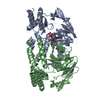
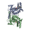
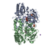
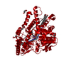
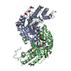
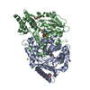
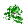
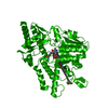
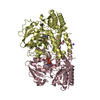
| Title | STRUCTURE OF PHOSPHOSERINE AMINOTRANSFERASE FROM ESCHERICHIA COLI | ||||||
 Components Components | PHOSPHOSERINE AMINOTRANSFERASE | ||||||
 Keywords Keywords | AMINOTRANSFERASE / L-SERINE BIOSYNTHESIS | ||||||
| Function / homology |  Function and homology information Function and homology information: / phosphoserine transaminase / O-phospho-L-serine:2-oxoglutarate transaminase activity / pyridoxal 5'-phosphate biosynthetic process / pyridoxine biosynthetic process / L-serine metabolic process / L-serine biosynthetic process / pyridoxal phosphate binding / protein homodimerization activity / cytosol / cytoplasm Similarity search - Function | ||||||
| Biological species |  | ||||||
| Method |  X-RAY DIFFRACTION / MIRAS / Resolution: 2.3 Å X-RAY DIFFRACTION / MIRAS / Resolution: 2.3 Å | ||||||
 Authors Authors | Hester, G. / Moser, M. / Jansonius, J.N. | ||||||
 Citation Citation | Journal: J.Mol.Biol. / Year: 1999 Title: Crystal structure of phosphoserine aminotransferase from Escherichia coli at 2.3 A resolution: comparison of the unligated enzyme and a complex with alpha-methyl-l-glutamate. Authors: Hester, G. / Stark, W. / Moser, M. / Kallen, J. / Markovic-Housley, Z. / Jansonius, J.N. #1: Journal: Enzymes Dependent on Pyridoxal Phosphate and Other Carbonyl Compounds as Cofactors: Proceedings of the 8Th International Symposium on Vitamin B6 and Carbonyl CatalysisYear: 1991 Title: The Three Dimensional Structure of Phosphoserine Aminotransferase from Escherichia Coli Authors: Stark, W. / Kallen, J. / Markovic-Housley, Z. / Fol, B. / Kania, M. / Jansonius, J.N. #2: Journal: Biochemistry of Vitamin B6: Proceedings of the 7Th International Congress on Chemical and Biological Aspects of Vitamin B6 Catalysis (in: Iub Symp. Ser., V.166)Year: 1987 Title: Crystallographic and Solution Studies on Phosphoserine Aminotransferase (Psat) from E. Coli Authors: Kallen, J. / Kania, M. / Markovic-Housley, Z. / Vincent, M.G. / Jansonius, J.N. | ||||||
| History |
|
- Structure visualization
| Structure viewer | Molecule: MolmilJmol/JSmol |
|---|
- Downloads & links
-Download
| PDBx/mmCIF format | 1bjn.cif.gz | 154.9 KB | Display | PDBx/mmCIF format |
|---|---|---|---|---|
| PDB format | pdb1bjn.ent.gz | 121.5 KB | Display | PDB format |
| PDBx/mmJSON format | 1bjn.json.gz | Tree view | PDBx/mmJSON format | |
| Others |  Other downloads Other downloads |
-Validation report
| Arichive directory | https://data.pdbj.org/pub/pdb/validation_reports/bj/1bjnftp://data.pdbj.org/pub/pdb/validation_reports/bj/1bjn | HTTPS FTP |
|---|
-Related structure data


-Links
 PDBj
PDBj- Assembly
| Deposited unit | 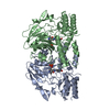
| ||||||||
|---|---|---|---|---|---|---|---|---|---|
| 1 |
| ||||||||
| Unit cell |
| ||||||||
| Noncrystallographic symmetry (NCS) | NCS oper: (Code: given Matrix: (0.45112, -0.73331, 0.50868), Vector: |
-Components
| #1: Protein | Mass: 39937.164 Da / Num. of mol.: 2 Source method: isolated from a genetically manipulated source Details: ONE COFACTOR, PYRIDOXAL PHOSPHATE (VITAMIN B6), IS PRESENT PER CHAIN Source: (gene. exp.) #2: Water | ChemComp-HOH / |  Mass: 18.015 Da / Num. of mol.: 241 / Source method: isolated from a natural source / Formula: H2O Mass: 18.015 Da / Num. of mol.: 241 / Source method: isolated from a natural source / Formula: H2O |
|---|
-Experimental details
-Experiment
| Experiment | Method: X-RAY DIFFRACTION / Number of used crystals: 1 |
|---|
- Sample preparation
| Crystal | Density Matthews: 2.7 Å3/Da / Density % sol: 52 % | ||||||||||||
|---|---|---|---|---|---|---|---|---|---|---|---|---|---|
| Crystal grow | Temperature: 277 K / Method: vapor diffusion, hanging drop Details: PROTEIN WAS CRYSTALLIZED AT 4-7 DEGREES CELSIUS BY HANGING DROP METHOD, USING PEG4000 AS A PRECIPITATING AGENT, BUFFERED WITH SODIUM ACETATE TO PH 7.2 IN THE DROP AND PH 5.6 IN THE RESERVOIR. ...Details: PROTEIN WAS CRYSTALLIZED AT 4-7 DEGREES CELSIUS BY HANGING DROP METHOD, USING PEG4000 AS A PRECIPITATING AGENT, BUFFERED WITH SODIUM ACETATE TO PH 7.2 IN THE DROP AND PH 5.6 IN THE RESERVOIR. THE PH-GRADIENT WAS ESSENTIAL FOR THE CRYSTALLIZATION., vapor diffusion - hanging drop, temperature 277K PH range: 5.6-7.2 | ||||||||||||
| Crystal grow | *PLUS Temperature: 4-7 ℃ / Method: vapor diffusion, hanging drop / Details: pH7.2 in the drop and pH5.6 in the reservoir | ||||||||||||
| Components of the solutions | *PLUS
|
-Data collection
| Diffraction | Mean temperature: 277 K |
|---|---|
| Diffraction source | Source: ROTATING ANODE / Type: ELLIOTT GX-20 / Wavelength: 1.5418 |
| Detector | Type: MARRESEARCH / Detector: IMAGE PLATE / Date: Jan 1, 1996 |
| Radiation | Monochromatic (M) / Laue (L): M / Scattering type: x-ray |
| Radiation wavelength | Wavelength: 1.5418 Å / Relative weight: 1 |
| Reflection | Resolution: 2.3→32.5 Å / Num. obs: 38831 / % possible obs: 98.2 % / Observed criterion σ(I): 0 / Redundancy: 4.5 % / Biso Wilson estimate: 29.8 Å2 / Rsym value: 0.083 / Net I/σ(I): 14.7 |
| Reflection shell | Resolution: 2.3→2.38 Å / Redundancy: 4.4 % / Mean I/σ(I) obs: 3.3 / Rsym value: 0.474 / % possible all: 99.9 |
| Reflection | *PLUS Rmerge(I) obs: 0.083 |
- Processing
| Software |
| ||||||||||||||||||||||||||||||||||||||||||||||||||||||||||||||||||||||||||||||||
|---|---|---|---|---|---|---|---|---|---|---|---|---|---|---|---|---|---|---|---|---|---|---|---|---|---|---|---|---|---|---|---|---|---|---|---|---|---|---|---|---|---|---|---|---|---|---|---|---|---|---|---|---|---|---|---|---|---|---|---|---|---|---|---|---|---|---|---|---|---|---|---|---|---|---|---|---|---|---|---|---|---|
| Refinement | Method to determine structure: MIRAS / Resolution: 2.3→32.5 Å / Rfactor Rfree error: 0.005 / Data cutoff high absF: 10000000 / Data cutoff low absF: 0.001 / Isotropic thermal model: RESTRAINED / Cross valid method: THROUGHOUT / σ(F): 0 Details: BULK SOLVENT MODEL USED. ATOMIC OCCUPANCIES OF RESIDUES WITH ALTERNATIVE CONFORMATIONS WERE REFINED DURING EACH REFINEMENT ROUND. IN THE LAST REFINEMENT ROUND THE OCCUPANCIES WERE FIXED, ...Details: BULK SOLVENT MODEL USED. ATOMIC OCCUPANCIES OF RESIDUES WITH ALTERNATIVE CONFORMATIONS WERE REFINED DURING EACH REFINEMENT ROUND. IN THE LAST REFINEMENT ROUND THE OCCUPANCIES WERE FIXED, RESULTING IN A TOTAL SUMMED OCCUPANCY FOR EACH RESIDUE OF 1.0.
| ||||||||||||||||||||||||||||||||||||||||||||||||||||||||||||||||||||||||||||||||
| Displacement parameters | Biso mean: 33 Å2 | ||||||||||||||||||||||||||||||||||||||||||||||||||||||||||||||||||||||||||||||||
| Refine analyze |
| ||||||||||||||||||||||||||||||||||||||||||||||||||||||||||||||||||||||||||||||||
| Refinement step | Cycle: LAST / Resolution: 2.3→32.5 Å
| ||||||||||||||||||||||||||||||||||||||||||||||||||||||||||||||||||||||||||||||||
| Refine LS restraints |
| ||||||||||||||||||||||||||||||||||||||||||||||||||||||||||||||||||||||||||||||||
| Refine LS restraints NCS | Rms dev Biso : 2.65 Å2 / Rms dev position: 0.54 Å / Weight Biso : 2.5 / Weight position: 100 | ||||||||||||||||||||||||||||||||||||||||||||||||||||||||||||||||||||||||||||||||
| LS refinement shell | Resolution: 2.3→2.35 Å / Rfactor Rfree error: 0.029 / Total num. of bins used: 15
| ||||||||||||||||||||||||||||||||||||||||||||||||||||||||||||||||||||||||||||||||
| Xplor file |
| ||||||||||||||||||||||||||||||||||||||||||||||||||||||||||||||||||||||||||||||||
| Software | *PLUS Name: X-PLOR / Version: 3.851 / Classification: refinement | ||||||||||||||||||||||||||||||||||||||||||||||||||||||||||||||||||||||||||||||||
| Refinement | *PLUS Rfactor all: 0.175 / Rfactor obs: 0.172 | ||||||||||||||||||||||||||||||||||||||||||||||||||||||||||||||||||||||||||||||||
| Solvent computation | *PLUS | ||||||||||||||||||||||||||||||||||||||||||||||||||||||||||||||||||||||||||||||||
| Displacement parameters | *PLUS | ||||||||||||||||||||||||||||||||||||||||||||||||||||||||||||||||||||||||||||||||
| Refine LS restraints | *PLUS
|
