 Movie
Movie Controller

[English] 日本語
 Yorodumi
- PDB-1bt4: PHOSPHOSERINE AMINOTRANSFERASE FROM BACILLUS CIRCULANS SUBSP. ALK... -
+ Open data

- Basic information
| Entry | Database: PDB / ID: 1bt4 | ||||||
|---|---|---|---|---|---|---|---|
| Title | PHOSPHOSERINE AMINOTRANSFERASE FROM BACILLUS CIRCULANS SUBSP. ALKALOPHILUS | ||||||
 Components Components | Phosphoserine aminotransferase | ||||||
 Keywords Keywords | TRANSFERASE / AMINOTRANSFERASE / PYRIDOXAL-5'-PHOSPHATE / PHOSPHOSERINE / ALKALIPHILIC | ||||||
| Function / homology |  Function and homology information Function and homology informationphosphoserine transaminase / O-phospho-L-serine:2-oxoglutarate transaminase activity / L-serine biosynthetic process / pyridoxal phosphate binding / cytoplasm Similarity search - Function | ||||||
| Biological species |  Bacillus circulans (bacteria) Bacillus circulans (bacteria) | ||||||
| Method |  X-RAY DIFFRACTION / MOLECULAR REPLACEMENT / Resolution: 2.3 Å X-RAY DIFFRACTION / MOLECULAR REPLACEMENT / Resolution: 2.3 Å | ||||||
 Authors Authors | Hester, G. / Luong, T.N. / Moser, M. / Jansonius, J.N. | ||||||
 Citation Citation | Journal: To be Published Title: The Crystal Structure of Phosphoserine Aminotransferase from Bacillus Circulans Subsp. Alkalophilus Authors: Hester, G. / Luong, T.N. / Moser, M. / Jansonius, J.N. #1: Journal: Protein Sci. / Year: 1996Title: Crystallization and Preliminary X-Ray Analysis of Phosphoserine Aminotransferase from Bacillus Circulans Subsp. Alkalophilus Authors: Moser, M. / Muller, R. / Battchikova, N. / Koivulehto, M. / Korpela, T. / Jansonius, J.N. #2: Journal: Biochim.Biophys.Acta / Year: 1996Title: Phosphoserine Aminotransferase from Bacillus Circulans Subsp. Alkalophilus: Purification, Gene Cloning and Sequencing Authors: Battchikova, N. / Himanen, J.-P. / Ahjolahti, M. / Korpela, T. | ||||||
| History |
|
- Structure visualization
| Structure viewer | Molecule: MolmilJmol/JSmol |
|---|
- Downloads & links
-Download
| PDBx/mmCIF format | 1bt4.cif.gz | 79.6 KB | Display | PDBx/mmCIF format |
|---|---|---|---|---|
| PDB format | pdb1bt4.ent.gz | 58.8 KB | Display | PDB format |
| PDBx/mmJSON format | 1bt4.json.gz | Tree view | PDBx/mmJSON format | |
| Others |  Other downloads Other downloads |
-Validation report
| Arichive directory | https://data.pdbj.org/pub/pdb/validation_reports/bt/1bt4ftp://data.pdbj.org/pub/pdb/validation_reports/bt/1bt4 | HTTPS FTP |
|---|
-Related structure data
| Related structure data |  1bjnS S: Starting model for refinement |
|---|---|
| Similar structure data |


-Links
 PDBj
PDBj- Assembly
| Deposited unit | 
| ||||||||
|---|---|---|---|---|---|---|---|---|---|
| 1 | 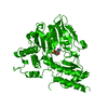
| ||||||||
| Unit cell |
|
-Components
| #1: Protein | Mass: 39834.895 Da / Num. of mol.: 1 / Fragment: ONE COMPLETE SUBUNIT / Mutation: K3E / Source method: isolated from a natural source Details: THE COFACTOR PYRIDOXAL-5'-PHOSPHATE IS COVALENTLY LINKED TO THE SIDE CHAIN OF LYS 197 Source: (natural) Bacillus circulans (bacteria) / Variant: ALKALOPHILUS / Strain: subsp. alkalophilus / References: UniProt: Q59196, phosphoserine transaminase | ||
|---|---|---|---|
| #2: Chemical | ChemComp-PLP /   Mass: 247.142 Da / Num. of mol.: 1 / Source method: obtained synthetically / Formula: C8H10NO6P Mass: 247.142 Da / Num. of mol.: 1 / Source method: obtained synthetically / Formula: C8H10NO6P | ||
| #3: Water | ChemComp-HOH /  Mass: 18.015 Da / Num. of mol.: 98 / Source method: isolated from a natural source / Formula: H2O Mass: 18.015 Da / Num. of mol.: 98 / Source method: isolated from a natural source / Formula: H2O | ||
| Nonpolymer details | PLP MOLECULE IS COVALENTLY| Sequence details | MET 1 IS CLEAVED OFF IN A POST-TRANSLATIO | |
-Experimental details
-Experiment
| Experiment | Method: X-RAY DIFFRACTION / Number of used crystals: 1 |
|---|
- Sample preparation
| Crystal | Density Matthews: 2.36 Å3/Da / Density % sol: 48 % |
|---|---|
| Crystal grow | Method: macroseeding / pH: 4.6 Details: CRYSTALLIZED AT ROOM TEMPERATURE FROM 0.1 M SODIUM ACETATE BUFFER, PH 4.6, 2-6% GLYCEROL, 2-4% PEG 20000, USING MACROSEEDING TECHNIQUES, macroseeding Temp details: room temp |
-Data collection
| Diffraction | Mean temperature: 277 K |
|---|---|
| Diffraction source | Source: ROTATING ANODE / Type: ENRAF-NONIUS / Wavelength: 1.5418 |
| Detector | Type: MARRESEARCH / Detector: IMAGE PLATE / Date: Jan 15, 1996 |
| Radiation | Protocol: SINGLE WAVELENGTH / Monochromatic (M) / Laue (L): M / Scattering type: x-ray |
| Radiation wavelength | Wavelength: 1.5418 Å / Relative weight: 1 |
| Reflection | Resolution: 2.3→29.3 Å / Num. obs: 16466 / % possible obs: 99.4 % / Redundancy: 3.6 % / Biso Wilson estimate: 19.3 Å2 / Rmerge(I) obs: 0.135 / Rsym value: 0.135 / Net I/σ(I): 7.3 |
| Reflection shell | Resolution: 2.3→2.38 Å / Redundancy: 3.4 % / Rmerge(I) obs: 0.219 / Mean I/σ(I) obs: 5 / Rsym value: 0.219 / % possible all: 96.4 |
- Processing
| Software |
| ||||||||||||||||||||||||||||||||||||||||||||||||||||||||||||
|---|---|---|---|---|---|---|---|---|---|---|---|---|---|---|---|---|---|---|---|---|---|---|---|---|---|---|---|---|---|---|---|---|---|---|---|---|---|---|---|---|---|---|---|---|---|---|---|---|---|---|---|---|---|---|---|---|---|---|---|---|---|
| Refinement | Method to determine structure: MOLECULAR REPLACEMENT Starting model: 1BJN Resolution: 2.3→29.3 Å / Rfactor Rfree error: 0.008 / Data cutoff high absF: 10000000 / Data cutoff low absF: 0.001 / Isotropic thermal model: GROUP / Cross valid method: THROUGHOUT / σ(F): 0 / Details: BULK SOLVENT MODEL USED
| ||||||||||||||||||||||||||||||||||||||||||||||||||||||||||||
| Displacement parameters | Biso mean: 20.7 Å2
| ||||||||||||||||||||||||||||||||||||||||||||||||||||||||||||
| Refine analyze |
| ||||||||||||||||||||||||||||||||||||||||||||||||||||||||||||
| Refinement step | Cycle: LAST / Resolution: 2.3→29.3 Å
| ||||||||||||||||||||||||||||||||||||||||||||||||||||||||||||
| Refine LS restraints |
| ||||||||||||||||||||||||||||||||||||||||||||||||||||||||||||
| LS refinement shell | Resolution: 2.3→2.35 Å / Rfactor Rfree error: 0.047 / Total num. of bins used: 15
| ||||||||||||||||||||||||||||||||||||||||||||||||||||||||||||
| Xplor file |
|
