Mass: 18.015 Da / Num. of mol.: 32 / Source method: isolated from a natural source / Formula: H2O
Has protein modification
Y
-
Experimental details
-
Experiment
Experiment
Method: X-RAY DIFFRACTION / Number of used crystals: 2
-
Sample preparation
Crystal
Density Matthews: 3.25 Å3/Da / Density % sol: 62.17 % Description: Bijvoet pairs were collected separately in the native data set. The averaged reflections were used for structure refinement. Mosaicity: 1.14 °
Crystal grow
Temperature: 293 K / Method: vapor diffusion, hanging drop / pH: 7.5 Details: 15%(w/v) PEG6000, 0.1M KCl, 0.1M HEPES-Na buffer, pH 7.5, VAPOR DIFFUSION, HANGING DROP, temperature 293.0K
-
Data collection
Diffraction
ID
Mean temperature (K)
Crystal-ID
1
100
1
2
100
1
Diffraction source
Source
Site
Beamline
ID
Wavelength (Å)
SYNCHROTRON
SPring-8
BL44XU
1
0.9
SYNCHROTRON
SPring-8
BL44XU
2
0.9791, 0.9794, 0.9642
Detector
Type
ID
Detector
Date
Details (eV)
Bruker DIP-6040
1
CCD
Feb 16, 2009
Horizontalfocusingmirror
Bruker DIP-6040
2
CCD
Jan 22, 2011
HORIZONTALFOCUSINGMIRROR
Radiation
ID
Monochromator
Protocol
Monochromatic (M) / Laue (L)
Scattering type
Wavelength-ID
1
Double-crystal monochromator
SINGLEWAVELENGTH
M
x-ray
1
2
DOUBLE-CRYSTAL MONOCHROMATOR
MAD
M
x-ray
2
Radiation wavelength
ID
Wavelength (Å)
Relative weight
1
0.9
1
2
0.9791
1
3
0.9794
1
4
0.9642
1
Reflection
Redundancy: 13.9 % / Av σ(I) over netI: 27.33 / Number: 99095 / Rmerge(I) obs: 0.137 / Χ2: 12.18 / D res high: 3.2 Å / D res low: 50 Å / Num. obs: 7141 / % possible obs: 99.2
Diffraction reflection shell
Highest resolution (Å)
Lowest resolution (Å)
% possible obs (%)
ID
Rmerge(I) obs
Chi squared
Redundancy
8.67
50
87.2
1
0.115
46.666
9.7
6.89
8.67
100
1
0.098
21.805
13.2
6.02
6.89
100
1
0.125
19.331
13.6
5.47
6.02
100
1
0.143
17.994
13.9
5.08
5.47
99.4
1
0.138
14.645
14
4.78
5.08
99.7
1
0.121
12.145
14
4.54
4.78
100
1
0.109
9.768
13.8
4.34
4.54
100
1
0.107
8.601
13.9
4.18
4.34
100
1
0.122
8.063
14.1
4.03
4.18
99.5
1
0.124
6.744
14.2
3.91
4.03
100
1
0.131
6.869
14.3
3.79
3.91
100
1
0.151
6.187
14.3
3.69
3.79
100
1
0.151
6.805
14.2
3.6
3.69
99.7
1
0.189
6.98
14.5
3.52
3.6
100
1
0.205
7.176
14.3
3.45
3.52
100
1
0.251
6.572
14.3
3.38
3.45
100
1
0.297
5.985
14.6
3.31
3.38
100
1
0.321
14.366
14.2
3.26
3.31
100
1
0.368
13.68
14.3
3.2
3.26
100
1
0.395
14.33
14.5
Reflection
Resolution: 2.7→300 Å / Num. obs: 20666 / % possible obs: 99.9 % / Observed criterion σ(I): 0 / Redundancy: 5.7 % / Biso Wilson estimate: 54 Å2 / Rmerge(I) obs: 0.103 / Χ2: 2.308 / Net I/σ(I): 18.8
Reflection shell
Resolution (Å)
Redundancy (%)
Rmerge(I) obs
Num. unique all
Χ2
Diffraction-ID
% possible all
2.7-2.8
5.7
0.332
2072
1.432
1
100
2.8-2.91
5.8
0.269
2041
1.55
1
100
2.91-3.04
5.7
0.227
2066
1.655
1
100
3.04-3.2
5.8
0.177
2060
1.823
1
100
3.2-3.4
5.7
0.137
2099
2.16
1
100
3.4-3.66
5.7
0.117
2013
2.639
1
100
3.66-4.03
5.6
0.097
2116
3.03
1
100
4.03-4.62
5.5
0.083
2066
3.139
1
100
4.62-5.82
5.6
0.077
2057
2.923
1
99.9
5.82-300
5.4
0.071
2076
2.847
1
99.5
-
Phasing
Phasing
Method: MAD
-
Processing
Software
Name
Version
Classification
NB
SCALEPACK
datascaling
SHARP
phasing
REFMAC
refinement
PDB_EXTRACT
3.1
dataextraction
HKL-2000
datacollection
HKL-2000
datareduction
Refinement
Method to determine structure: MAD / Resolution: 2.71→56.27 Å / Cor.coef. Fo:Fc: 0.929 / Cor.coef. Fo:Fc free: 0.904 / WRfactor Rfree: 0.269 / WRfactor Rwork: 0.2361 / Occupancy max: 1 / Occupancy min: 1 / FOM work R set: 0.7971 / SU B: 27.035 / SU ML: 0.244 / SU R Cruickshank DPI: 0.4528 / SU Rfree: 0.2899 / Cross valid method: THROUGHOUT / σ(F): 0 / ESU R Free: 0.29 / Stereochemistry target values: MAXIMUM LIKELIHOOD
Rfactor
Num. reflection
% reflection
Selection details
Rfree
0.2525
539
4.8 %
RANDOM
Rwork
0.2182
-
-
-
obs
0.2199
10718
99.63 %
-
Solvent computation
Ion probe radii: 0.8 Å / Shrinkage radii: 0.8 Å / VDW probe radii: 1.4 Å / Solvent model: BABINET MODEL WITH MASK
In the structure databanks used in Yorodumi, some data are registered as the other names, "COVID-19 virus" and "2019-nCoV". Here are the details of the virus and the list of structure data.
Jan 31, 2019. EMDB accession codes are about to change! (news from PDBe EMDB page)
EMDB accession codes are about to change! (news from PDBe EMDB page)
The allocation of 4 digits for EMDB accession codes will soon come to an end. Whilst these codes will remain in use, new EMDB accession codes will include an additional digit and will expand incrementally as the available range of codes is exhausted. The current 4-digit format prefixed with “EMD-” (i.e. EMD-XXXX) will advance to a 5-digit format (i.e. EMD-XXXXX), and so on. It is currently estimated that the 4-digit codes will be depleted around Spring 2019, at which point the 5-digit format will come into force.
The EM Navigator/Yorodumi systems omit the EMD- prefix.
Related info.:Q: What is EMD? / ID/Accession-code notation in Yorodumi/EM Navigator
Yorodumi is a browser for structure data from EMDB, PDB, SASBDB, etc.
This page is also the successor to EM Navigator detail page, and also detail information page/front-end page for Omokage search.
The word "yorodu" (or yorozu) is an old Japanese word meaning "ten thousand". "mi" (miru) is to see.
Related info.:EMDB / PDB / SASBDB / Comparison of 3 databanks / Yorodumi Search / Aug 31, 2016. New EM Navigator & Yorodumi / Yorodumi Papers / Jmol/JSmol / Function and homology information / Changes in new EM Navigator and Yorodumi
 Movie
Movie Controller

 Yorodumi
 Open data
 Basic information
 Components
 Keywords
 Function and homology information
 Homo sapiens (human)
 X-RAY DIFFRACTION /
  Authors
 Citation
 Structure visualization
 Downloads & links
 Other downloads
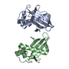


 PDBj
PDBj


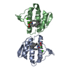
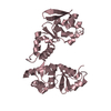
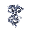
 Assembly

 Mass: 18.015 Da / Num. of mol.: 32 / Source method: isolated from a natural source / Formula: H2O
 Sample preparation
 Processing