 Movie
Movie Controller

[English] 日本語
 Yorodumi
- PDB-1l5t: Crystal Structure of a Domain-Opened Mutant (R121D) of the Human ... -
+ Open data

- Basic information
| Entry | Database: PDB / ID: 1l5t | ||||||
|---|---|---|---|---|---|---|---|
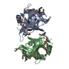
| Title | Crystal Structure of a Domain-Opened Mutant (R121D) of the Human Lactoferrin N-lobe Refined From a Merohedrally-Twinned Crystal Form. | ||||||
 Components Components | lactoferrin | ||||||
 Keywords Keywords | METAL TRANSPORT / IRON TRANSPORT / GLYCOPROTEIN / LACTOFERRIN / N-LOBE / IRON-RELEASE / TWINNING | ||||||
| Function / homology |  Function and homology information Function and homology informationmembrane destabilizing activity / host-mediated suppression of viral proces / Mtb iron assimilation by chelation / phagocytic vesicle lumen / Metal sequestration by antimicrobial proteins / negative regulation of viral process / negative regulation of tumor necrosis factor (ligand) superfamily member 11 production / positive regulation of toll-like receptor 4 signaling pathway / negative regulation of single-species biofilm formation in or on host organism / positive regulation of bone mineralization involved in bone maturation ...membrane destabilizing activity / host-mediated suppression of viral proces / Mtb iron assimilation by chelation / phagocytic vesicle lumen / Metal sequestration by antimicrobial proteins / negative regulation of viral process / negative regulation of tumor necrosis factor (ligand) superfamily member 11 production / positive regulation of toll-like receptor 4 signaling pathway / negative regulation of single-species biofilm formation in or on host organism / positive regulation of bone mineralization involved in bone maturation / negative regulation of ATP-dependent activity / negative regulation of osteoclast development / specific granule / negative regulation of lipopolysaccharide-mediated signaling pathway / positive regulation of chondrocyte proliferation / antifungal humoral response / regulation of tumor necrosis factor production / bone morphogenesis / Antimicrobial peptides / positive regulation of osteoblast proliferation / negative regulation of viral genome replication / positive regulation of protein serine/threonine kinase activity / humoral immune response / Hydrolases; Acting on peptide bonds (peptidases); Serine endopeptidases / positive regulation of osteoblast differentiation / cysteine-type endopeptidase inhibitor activity / : / regulation of cytokine production / secretory granule / ossification / protein serine/threonine kinase activator activity / innate immune response in mucosa / iron ion transport / lipopolysaccharide binding / recycling endosome / specific granule lumen / tertiary granule lumen / antimicrobial humoral immune response mediated by antimicrobial peptide / heparin binding / antibacterial humoral response / killing of cells of another organism / defense response to Gram-negative bacterium / early endosome / positive regulation of canonical NF-kappaB signal transduction / iron ion binding / Amyloid fiber formation / serine-type endopeptidase activity / Neutrophil degranulation / negative regulation of apoptotic process / cell surface / protein-containing complex / proteolysis / : / DNA binding / extracellular exosome / extracellular region / nucleus / plasma membrane / cytoplasm Similarity search - Function | ||||||
| Biological species |  Homo sapiens (human) Homo sapiens (human) | ||||||
| Method |  X-RAY DIFFRACTION / MOLECULAR REPLACEMENT / Resolution: 3 Å X-RAY DIFFRACTION / MOLECULAR REPLACEMENT / Resolution: 3 Å | ||||||
 Authors Authors | Jameson, G.B. / Anderson, B.F. / Breyer, W.A. / Tweedie, J.W. / Baker, E.N. | ||||||
 Citation Citation | Journal: Acta Crystallogr.,Sect.D / Year: 2002 Title: Structure of a domain-opened mutant (R121D) of the human lactoferrin N-lobe refined from a merohedrally twinned crystal form. Authors: Jameson, G.B. / Anderson, B.F. / Breyer, W.A. / Day, C.L. / Tweedie, J.W. / Baker, E.N. #1: Journal: Acta Crystallogr.,Sect.D / Year: 1999Title: On the Molecular-Replacement Problem in the Presence of Merohedral Twinning: Structure of the N-Terminal Half-Molecule of Human Lactoferrin Authors: Breyer, W.A. / Kingston, R.L. / Anderson, B.F. / Baker, E.N. | ||||||
| History |
|
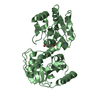
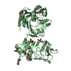
- Structure visualization
| Structure viewer | Molecule: MolmilJmol/JSmol |
|---|
- Downloads & links
-Download
| PDBx/mmCIF format | 1l5t.cif.gz | 137.7 KB | Display | PDBx/mmCIF format |
|---|---|---|---|---|
| PDB format | pdb1l5t.ent.gz | 108.3 KB | Display | PDB format |
| PDBx/mmJSON format | 1l5t.json.gz | Tree view | PDBx/mmJSON format | |
| Others |  Other downloads Other downloads |
-Validation report
| Arichive directory | https://data.pdbj.org/pub/pdb/validation_reports/l5/1l5tftp://data.pdbj.org/pub/pdb/validation_reports/l5/1l5t | HTTPS FTP |
|---|
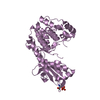
-Related structure data
| Related structure data |  1lctS S: Starting model for refinement |
|---|---|
| Similar structure data |


-Links
 PDBj
PDBj


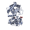
- Assembly
| Deposited unit | 
| ||||||||
|---|---|---|---|---|---|---|---|---|---|
| 1 | 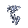
| ||||||||
| 2 | 
| ||||||||
| Unit cell |
|
-Components
| #1: Protein | Mass: 36921.875 Da / Num. of mol.: 2 / Fragment: RESIDUES 21-352 / Mutation: R121D Source method: isolated from a genetically manipulated source Source: (gene. exp.) Homo sapiens (human) / Plasmid: pnut / Cell line (production host): BHK / Production host:   Cricetulus griseus (Chinese hamster) / References: UniProt: P02788, EC: 1.16.1.2 Cricetulus griseus (Chinese hamster) / References: UniProt: P02788, EC: 1.16.1.2#2: Water | ChemComp-HOH / |  Mass: 18.015 Da / Num. of mol.: 127 / Source method: isolated from a natural source / Formula: H2O Mass: 18.015 Da / Num. of mol.: 127 / Source method: isolated from a natural source / Formula: H2OHas protein modification | Y | |
|---|
-Experimental details
-Experiment
| Experiment | Method: X-RAY DIFFRACTION / Number of used crystals: 1 |
|---|
- Sample preparation
| Crystal | Density Matthews: 4.35 Å3/Da / Density % sol: 71.7 % | |||||||||||||||||||||||||||||||||||||||||||||||||
|---|---|---|---|---|---|---|---|---|---|---|---|---|---|---|---|---|---|---|---|---|---|---|---|---|---|---|---|---|---|---|---|---|---|---|---|---|---|---|---|---|---|---|---|---|---|---|---|---|---|---|
| Crystal grow | Temperature: 298 K / Method: microdialysis / pH: 8 Details: concentrated solution of the protein (50-80 mg mL-1), 0.01 M Tris-HCl, pH 8.0, 12% (v/v) isopropanol, MICRODIALYSIS, temperature 298K | |||||||||||||||||||||||||||||||||||||||||||||||||
| Crystal grow | *PLUS | |||||||||||||||||||||||||||||||||||||||||||||||||
| Components of the solutions | *PLUS
|
-Data collection
| Diffraction | Mean temperature: 295 K |
|---|---|
| Diffraction source | Source: ROTATING ANODE / Type: RIGAKU RU200 / Wavelength: 1.5418 / Wavelength: 1.5418 Å |
| Detector | Type: RIGAKU RAXIS IIC / Detector: IMAGE PLATE / Date: Mar 1, 1994 / Details: graphite monochromator |
| Radiation | Monochromator: GRAPHITE / Protocol: SINGLE WAVELENGTH / Monochromatic (M) / Laue (L): M / Scattering type: x-ray |
| Radiation wavelength | Wavelength: 1.5418 Å / Relative weight: 1 |
| Reflection | Resolution: 3→50 Å / Num. obs: 23431 / % possible obs: 96 % / Observed criterion σ(F): 4 / Redundancy: 2.8 % / Rmerge(I) obs: 0.115 / Net I/σ(I): 6.1 |
| Reflection shell | Resolution: 3→3.11 Å / Redundancy: 2.4 % / Rmerge(I) obs: 0.418 / Mean I/σ(I) obs: 1.8 / Rsym value: 0.418 / % possible all: 93 |
| Reflection | *PLUS Lowest resolution: 30 Å / Num. obs: 23882 / % possible obs: 96 % |
| Reflection shell | *PLUS Highest resolution: 3 Å / % possible obs: 93 % |
- Processing
| Software |
| |||||||||||||||||||||||||||||||||
|---|---|---|---|---|---|---|---|---|---|---|---|---|---|---|---|---|---|---|---|---|---|---|---|---|---|---|---|---|---|---|---|---|---|---|
| Refinement | Method to determine structure: MOLECULAR REPLACEMENT Starting model: pdb entry 1lct Resolution: 3→10 Å / Num. parameters: 19883 / Num. restraintsaints: 27798 Isotropic thermal model: INDIVIDUAL, WITH TIGHT ADJACENCY AND NCS RESTRAINTS, AND IDENTITY CONSTRAINTS ON NEARLY CHEMICALLY EQUIVALENT REDIDUES (E.G. OE1 AND OE2 OF GLU) Cross valid method: THROUGHOUT / σ(F): 4 / σ(I): 2 / Stereochemistry target values: Engh & Huber Details: WEIGHTED CONJUGATE-GRADIENT REFINEMENT ON F-SQUARED; HEMIHEDRAL TWINNING TO GIVE PSEUDO-HEXAGONAL (6/M) DIFFRACTION SYMMETRY; TWIN RATIO=0.44035:0.54965. WITH REGARDS TO THE TORSION ANGLES ...Details: WEIGHTED CONJUGATE-GRADIENT REFINEMENT ON F-SQUARED; HEMIHEDRAL TWINNING TO GIVE PSEUDO-HEXAGONAL (6/M) DIFFRACTION SYMMETRY; TWIN RATIO=0.44035:0.54965. WITH REGARDS TO THE TORSION ANGLES OF LEU 299, THE RESIDUE IS PART OF A CONSERVED GAMMA TURN.
| |||||||||||||||||||||||||||||||||
| Solvent computation | Solvent model: MOEWS & KRETSINGER, 1975 K(SOLV)=0.551302, B(SOLV)=2.7660 Bsol: 2.766 Å2 / ksol: 0.551302 e/Å3 | |||||||||||||||||||||||||||||||||
| Displacement parameters | Biso mean: 31.1 Å2 | |||||||||||||||||||||||||||||||||
| Refine analyze | Num. disordered residues: 0 / Occupancy sum hydrogen: 0 / Occupancy sum non hydrogen: 5241 | |||||||||||||||||||||||||||||||||
| Refinement step | Cycle: LAST / Resolution: 3→10 Å
| |||||||||||||||||||||||||||||||||
| Refine LS restraints |
| |||||||||||||||||||||||||||||||||
| LS refinement shell | Resolution: 3→3.11 Å
| |||||||||||||||||||||||||||||||||
| Software | *PLUS Name: SHELXL / Version: 97 / Classification: refinement | |||||||||||||||||||||||||||||||||
| Refinement | *PLUS Highest resolution: 3 Å / Lowest resolution: 20 Å / Num. reflection obs: 22636 | |||||||||||||||||||||||||||||||||
| Solvent computation | *PLUS | |||||||||||||||||||||||||||||||||
| Displacement parameters | *PLUS |
