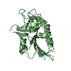
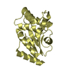
SHEET DETERMINATION METHOD: DSSP THE SHEETS PRESENTED AS "AD BC" IN EACH CHAIN ON SHEET RECORDS ... SHEET DETERMINATION METHOD: DSSP THE SHEETS PRESENTED AS "AD BC" IN EACH CHAIN ON SHEET RECORDS BELOW IS ACTUALLY AN 6-STRANDED BARREL THIS IS REPRESENTED BY A 7-STRANDED SHEET IN WHICH THE FIRST AND LAST STRANDS ARE IDENTICAL.
Mass: 18.015 Da / Num. of mol.: 98 / Source method: isolated from a natural source / Formula: H2O
Compound details
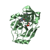
ENGINEERED RESIDUE IN CHAIN A B, CYS 95 TO LYS ENGINEERED RESIDUE IN CHAIN A B, CYS 142 TO SER ...ENGINEERED RESIDUE IN CHAIN A B, CYS 95 TO LYS ENGINEERED RESIDUE IN CHAIN A B, CYS 142 TO SER ENGINEERED RESIDUE IN CHAIN A B, CYS 163 TO ALA THIS PROTEIN CONTAINS THREE SUBSTITUTED CYS RESIDUES (C95K, C142S) TO ENHANCE SOLUBILITY AND ONE (C163A) WHICH SUBSTITUTES THE ACTIVE SITE NUCLEOPHILE AND THEREFORE INACTIVATES THE ENZYME.
Has protein modification
Y
Sequence details
CRYSTALLISED SEQUENCE CONTAINS AN ADDITIONAL GLY AT N-TERM (RESIDUE NUMBER -1). ALSO CONTAINS ...CRYSTALLISED SEQUENCE CONTAINS AN ADDITIONAL GLY AT N-TERM (RESIDUE NUMBER -1). ALSO CONTAINS TRUNCATED C-TERMINUS (BY 6 RESIDUES). DATABASE ENTRY CONTAINS A PROTEIN SEQUENCE ERROR. THE SEQUENCE 78-RTGHALRRGTHW-89 IN THE DATABASE ENTRY SHOULD BE REPLACED BY 78-GQDMLSDAALMV-89. THIS IS NOT EXPLICITLY CORRECTED IN THE SEQUENCE CONFLICTS GIVEN BELOW (CONFINED TO ENGINEERED MUTATIONS). THE FOLLOWING RESIDUES ARE CONFLICTS BETWEEN THE PROTEIN AND THE UNIPROT ENTRY, BUT SINCE THEY ARE NOT OBSERVED IN THE STRUCTURE, THEY CANNOT BE GIVEN SEQADV RECORDS: B78 (GLY), B79 (GLN), B80 (ASP), B81 (MSE) RESIDUES A142 (SER) AND B142 (SER) ARE ENGINEERED MUTATIONS, BUT THEY ARE NOT OBSERVED AND ARE NOT THEREFORE RECORDED WITH SEQADV RECORDS.
-
Experimental details
-
Experiment
Experiment
Method: X-RAY DIFFRACTION / Number of used crystals: 1
-
Sample preparation
Crystal
Density Matthews: 1.83 Å3/Da / Density % sol: 32 %
In the structure databanks used in Yorodumi, some data are registered as the other names, "COVID-19 virus" and "2019-nCoV". Here are the details of the virus and the list of structure data.
Jan 31, 2019. EMDB accession codes are about to change! (news from PDBe EMDB page)
EMDB accession codes are about to change! (news from PDBe EMDB page)
The allocation of 4 digits for EMDB accession codes will soon come to an end. Whilst these codes will remain in use, new EMDB accession codes will include an additional digit and will expand incrementally as the available range of codes is exhausted. The current 4-digit format prefixed with “EMD-” (i.e. EMD-XXXX) will advance to a 5-digit format (i.e. EMD-XXXXX), and so on. It is currently estimated that the 4-digit codes will be depleted around Spring 2019, at which point the 5-digit format will come into force.
The EM Navigator/Yorodumi systems omit the EMD- prefix.
Related info.:Q: What is EMD? / ID/Accession-code notation in Yorodumi/EM Navigator
Yorodumi is a browser for structure data from EMDB, PDB, SASBDB, etc.
This page is also the successor to EM Navigator detail page, and also detail information page/front-end page for Omokage search.
The word "yorodu" (or yorozu) is an old Japanese word meaning "ten thousand". "mi" (miru) is to see.
Related info.:EMDB / PDB / SASBDB / Comparison of 3 databanks / Yorodumi Search / Aug 31, 2016. New EM Navigator & Yorodumi / Yorodumi Papers / Jmol/JSmol / Function and homology information / Changes in new EM Navigator and Yorodumi
 Movie
Movie Controller


 Open data
 Basic information
 Components
 Keywords
 Function and homology information
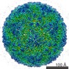 FOOT-AND-MOUTH DISEASE VIRUS
 X-RAY DIFFRACTION /
  Authors
 Citation
 Structure visualization
 Downloads & links
 Other downloads
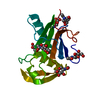


 PDBj
PDBj

 Assembly

 Mass: 18.015 Da / Num. of mol.: 98 / Source method: isolated from a natural source / Formula: H2O
 Sample preparation
 / Beamline: BM14 / Wavelength: 0.98
  Processing