 Movie
Movie Controller


+ Open data

- Basic information
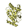
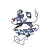
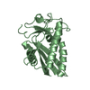
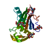
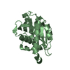
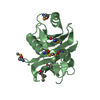
| Entry | Database: PDB / ID: 3s6u | ||||||
|---|---|---|---|---|---|---|---|
| Title | Caclcium-bound Ac-ASP-7 | ||||||
 Components Components | Ac-ASP-7 | ||||||
 Keywords Keywords | UNKNOWN FUNCTION / PR domain / CAP domain / SCP/TAPS | ||||||
| Function / homology | Pathogenesis-related Protein p14a / CAP / 3-Layer(aba) Sandwich / Alpha Beta Function and homology information Function and homology information | ||||||
| Biological species |  Ancylostoma caninum (dog hookworm) Ancylostoma caninum (dog hookworm) | ||||||
| Method |  X-RAY DIFFRACTION / SYNCHROTRON / MOLECULAR REPLACEMENT / Resolution: 2.7 Å X-RAY DIFFRACTION / SYNCHROTRON / MOLECULAR REPLACEMENT / Resolution: 2.7 Å | ||||||
 Authors Authors | Osman, A. / Wang, C.K. / Winter, A. / Loukas, A. / Tribolet, L. / Gasser, R. / Hofmann, A. | ||||||
 Citation Citation | Journal: Biotechnol Adv Title: Hookworm SCP/TAPS protein structure--A key to understanding host-parasite interactions and developing new interventions. Authors: Osman, A. / Wang, C.K. / Winter, A. / Loukas, A. / Tribolet, L. / Gasser, R.B. / Hofmann, A. | ||||||
| History |
|
- Structure visualization
| Structure viewer | Molecule: MolmilJmol/JSmol |
|---|
- Downloads & links
-Download
| PDBx/mmCIF format | 3s6u.cif.gz | 82.9 KB | Display | PDBx/mmCIF format |
|---|---|---|---|---|
| PDB format | pdb3s6u.ent.gz | 61.8 KB | Display | PDB format |
| PDBx/mmJSON format | 3s6u.json.gz | Tree view | PDBx/mmJSON format | |
| Others |  Other downloads Other downloads |
-Validation report
| Arichive directory | https://data.pdbj.org/pub/pdb/validation_reports/s6/3s6uftp://data.pdbj.org/pub/pdb/validation_reports/s6/3s6u | HTTPS FTP |
|---|
-Related structure data
| Related structure data | 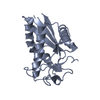 3s6sSC  3s6vC S: Starting model for refinement C: citing same article ( |
|---|---|
| Similar structure data |

-Links
 PDBj
PDBj- Assembly
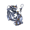
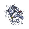
| Deposited unit | 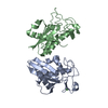
| ||||||||
|---|---|---|---|---|---|---|---|---|---|
| 1 | 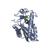
| ||||||||
| 2 | 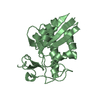
| ||||||||
| Unit cell |
|
-Components
| #1: Protein | Mass: 23511.436 Da / Num. of mol.: 2 Source method: isolated from a genetically manipulated source Source: (gene. exp.) Ancylostoma caninum (dog hookworm) / Production host:  Pichia pastoris (fungus) Pichia pastoris (fungus)#2: Chemical | ChemComp-CA / |   Mass: 40.078 Da / Num. of mol.: 1 / Source method: obtained synthetically / Formula: Ca Mass: 40.078 Da / Num. of mol.: 1 / Source method: obtained synthetically / Formula: Ca#3: Water | ChemComp-HOH / |  Mass: 18.015 Da / Num. of mol.: 100 / Source method: isolated from a natural source / Formula: H2O Mass: 18.015 Da / Num. of mol.: 100 / Source method: isolated from a natural source / Formula: H2OHas protein modification | Y | Sequence details | AS PER THE THE AUTHORS THERE IS AN UNEXPLAINED SEQUENCE MISMATCH BETWEEN THE GENBANK SEQUENCE AND ...AS PER THE THE AUTHORS THERE IS AN UNEXPLAINE | |
|---|
-Experimental details
-Experiment
| Experiment | Method: X-RAY DIFFRACTION / Number of used crystals: 1 |
|---|
- Sample preparation
| Crystal | Density Matthews: 2.35 Å3/Da / Density % sol: 47.63 % |
|---|---|
| Crystal grow | Temperature: 289 K / Method: vapor diffusion, hanging drop / pH: 7 Details: 20% MPD, 0.1 M TRIS, pH 7, VAPOR DIFFUSION, HANGING DROP, temperature 289K |
-Data collection
| Diffraction | Mean temperature: 100 K |
|---|---|
| Diffraction source | Source: SYNCHROTRON / Site: Australian Synchrotron  / Beamline: MX1 / Wavelength: 1.3778 Å / Beamline: MX1 / Wavelength: 1.3778 Å |
| Detector | Type: ADSC QUANTUM 315 / Detector: CCD / Date: Mar 29, 2011 |
| Radiation | Protocol: SINGLE WAVELENGTH / Monochromatic (M) / Laue (L): M / Scattering type: x-ray |
| Radiation wavelength | Wavelength: 1.3778 Å / Relative weight: 1 |
| Reflection | Resolution: 2.7→61 Å / Num. obs: 23262 / % possible obs: 99.2 % / Redundancy: 14.1 % / Biso Wilson estimate: 54.2 Å2 / Rsym value: 0.063 |
| Reflection shell | Resolution: 2.7→2.85 Å / Redundancy: 14.5 % / Mean I/σ(I) obs: 14.9 / Num. unique all: 1777 / Rsym value: 0.169 / % possible all: 98.6 |
- Processing
| Software |
| |||||||||||||||||||||||||||||||||||||||||||||||||||||||||||||||
|---|---|---|---|---|---|---|---|---|---|---|---|---|---|---|---|---|---|---|---|---|---|---|---|---|---|---|---|---|---|---|---|---|---|---|---|---|---|---|---|---|---|---|---|---|---|---|---|---|---|---|---|---|---|---|---|---|---|---|---|---|---|---|---|---|
| Refinement | Method to determine structure: MOLECULAR REPLACEMENT Starting model: PDB entry 3S6S Resolution: 2.7→24.78 Å / SU ML: 0.35 / σ(F): 1.6 / Phase error: 23.23 / Stereochemistry target values: ML
| |||||||||||||||||||||||||||||||||||||||||||||||||||||||||||||||
| Solvent computation | Shrinkage radii: 0.83 Å / VDW probe radii: 1.1 Å / Solvent model: FLAT BULK SOLVENT MODEL / Bsol: 34.588 Å2 / ksol: 0.348 e/Å3 | |||||||||||||||||||||||||||||||||||||||||||||||||||||||||||||||
| Displacement parameters |
| |||||||||||||||||||||||||||||||||||||||||||||||||||||||||||||||
| Refinement step | Cycle: LAST / Resolution: 2.7→24.78 Å
| |||||||||||||||||||||||||||||||||||||||||||||||||||||||||||||||
| Refine LS restraints |
| |||||||||||||||||||||||||||||||||||||||||||||||||||||||||||||||
| LS refinement shell |
|
