 Movie
Movie Controller

[English] 日本語
 Yorodumi
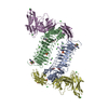
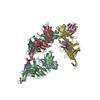
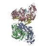
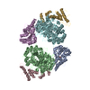
- PDB-5xwu: Crystal structure of PTPdelta Ig1-Ig3 in complex with SALM2 LRR-Ig -
+ Open data

- Basic information
| Entry | Database: PDB / ID: 5xwu | |||||||||
|---|---|---|---|---|---|---|---|---|---|---|
| Title | Crystal structure of PTPdelta Ig1-Ig3 in complex with SALM2 LRR-Ig | |||||||||
 Components Components |
| |||||||||
 Keywords Keywords | CELL ADHESION / synaptic orgnizers | |||||||||
| Function / homology |  Function and homology information Function and homology informationtrans-synaptic signaling / Receptor-type tyrosine-protein phosphatases / trans-synaptic signaling by trans-synaptic complex / Synaptic adhesion-like molecules / presynaptic membrane assembly / synaptic membrane adhesion / presynapse assembly / regulation of postsynaptic density assembly / negative regulation of receptor signaling pathway via JAK-STAT / positive regulation of dendritic spine morphogenesis ...trans-synaptic signaling / Receptor-type tyrosine-protein phosphatases / trans-synaptic signaling by trans-synaptic complex / Synaptic adhesion-like molecules / presynaptic membrane assembly / synaptic membrane adhesion / presynapse assembly / regulation of postsynaptic density assembly / negative regulation of receptor signaling pathway via JAK-STAT / positive regulation of dendritic spine morphogenesis / positive regulation of dendrite morphogenesis / positive regulation of synapse assembly / heterophilic cell-cell adhesion / regulation of immune response / protein-tyrosine-phosphatase / presynaptic active zone membrane / protein tyrosine phosphatase activity / cell adhesion molecule binding / hippocampal mossy fiber to CA3 synapse / postsynaptic density membrane / modulation of chemical synaptic transmission / Schaffer collateral - CA1 synapse / neuron differentiation / nervous system development / presynaptic membrane / signaling receptor binding / glutamatergic synapse / cell surface / signal transduction Similarity search - Function | |||||||||
| Biological species |  | |||||||||
| Method |  X-RAY DIFFRACTION / SYNCHROTRON / Resolution: 3.162 Å X-RAY DIFFRACTION / SYNCHROTRON / Resolution: 3.162 Å | |||||||||
 Authors Authors | Goto-Ito, S. / Yamagata, A. / Sato, Y. / Fukai, S. | |||||||||
 Citation Citation | Journal: Nat Commun / Year: 2018 Title: Structural basis of trans-synaptic interactions between PTP delta and SALMs for inducing synapse formation. Authors: Goto-Ito, S. / Yamagata, A. / Sato, Y. / Uemura, T. / Shiroshima, T. / Maeda, A. / Imai, A. / Mori, H. / Yoshida, T. / Fukai, S. | |||||||||
| History |
|
- Structure visualization
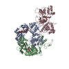
| Structure viewer | Molecule: MolmilJmol/JSmol |
|---|
- Downloads & links
-Download
| PDBx/mmCIF format | 5xwu.cif.gz | 259.7 KB | Display | PDBx/mmCIF format |
|---|---|---|---|---|
| PDB format | pdb5xwu.ent.gz | 206.9 KB | Display | PDB format |
| PDBx/mmJSON format | 5xwu.json.gz | Tree view | PDBx/mmJSON format | |
| Others |  Other downloads Other downloads |
-Validation report
| Arichive directory | https://data.pdbj.org/pub/pdb/validation_reports/xw/5xwuftp://data.pdbj.org/pub/pdb/validation_reports/xw/5xwu | HTTPS FTP |
|---|
-Related structure data


-Links
 PDBj
PDBj


- Assembly
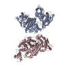
| Deposited unit | 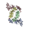
| ||||||||
|---|---|---|---|---|---|---|---|---|---|
| 1 |
| ||||||||
| Unit cell |
|
-Components
| #1: Protein | Mass: 34432.973 Da / Num. of mol.: 2 / Fragment: UNP residues 20-321 Source method: isolated from a genetically manipulated source Source: (gene. exp.)  Homo sapiens (human) / References: UniProt: Q64487, protein-tyrosine-phosphatase Homo sapiens (human) / References: UniProt: Q64487, protein-tyrosine-phosphatase#2: Protein | Mass: 40254.949 Da / Num. of mol.: 2 / Fragment: UNP residues 32-390 Source method: isolated from a genetically manipulated source Source: (gene. exp.) Homo sapiens (human) / References: UniProt: Q2WF71#3: Polysaccharide | Source method: isolated from a genetically manipulated source #4: Sugar | ChemComp-NAG /   Type: D-saccharide, beta linking / Mass: 221.208 Da / Num. of mol.: 4 Type: D-saccharide, beta linking / Mass: 221.208 Da / Num. of mol.: 4Source method: isolated from a genetically manipulated source Formula: C8H15NO6 #5: Chemical |   Mass: 195.237 Da / Num. of mol.: 2 / Source method: obtained synthetically / Formula: C6H13NO4S / Comment: pH buffer*YM Mass: 195.237 Da / Num. of mol.: 2 / Source method: obtained synthetically / Formula: C6H13NO4S / Comment: pH buffer*YMHas protein modification | Y | |
|---|
-Experimental details
-Experiment
| Experiment | Method: X-RAY DIFFRACTION / Number of used crystals: 1 |
|---|
- Sample preparation
| Crystal | Density Matthews: 4.04 Å3/Da / Density % sol: 69.58 % |
|---|---|
| Crystal grow | Temperature: 293 K / Method: vapor diffusion, sitting drop / Details: 7% PEG 20000, 0.1M MES (pH 6.0) |
-Data collection
| Diffraction | Mean temperature: 100 K |
|---|---|
| Diffraction source | Source: SYNCHROTRON / Site: SPring-8  / Beamline: BL41XU / Wavelength: 1 Å / Beamline: BL41XU / Wavelength: 1 Å |
| Detector | Type: DECTRIS PILATUS3 S 6M / Detector: PIXEL / Date: Jul 11, 2016 |
| Radiation | Protocol: SINGLE WAVELENGTH / Monochromatic (M) / Laue (L): M / Scattering type: x-ray |
| Radiation wavelength | Wavelength: 1 Å / Relative weight: 1 |
| Reflection | Resolution: 3.18→50 Å / Num. obs: 41658 / % possible obs: 99.8 % / Redundancy: 9.6 % / Net I/σ(I): 7.48 |
- Processing
| Software |
| |||||||||||||||||||||||||||||||||||||||||||||||||||||||||||||||||||||||||||||||||||||||||||||||||||||||||
|---|---|---|---|---|---|---|---|---|---|---|---|---|---|---|---|---|---|---|---|---|---|---|---|---|---|---|---|---|---|---|---|---|---|---|---|---|---|---|---|---|---|---|---|---|---|---|---|---|---|---|---|---|---|---|---|---|---|---|---|---|---|---|---|---|---|---|---|---|---|---|---|---|---|---|---|---|---|---|---|---|---|---|---|---|---|---|---|---|---|---|---|---|---|---|---|---|---|---|---|---|---|---|---|---|---|---|
| Refinement | Resolution: 3.162→48.71 Å / SU ML: 0.46 / Cross valid method: FREE R-VALUE / σ(F): 1.35 / Phase error: 26.84 / Stereochemistry target values: ML
| |||||||||||||||||||||||||||||||||||||||||||||||||||||||||||||||||||||||||||||||||||||||||||||||||||||||||
| Solvent computation | Shrinkage radii: 0.9 Å / VDW probe radii: 1.11 Å / Solvent model: FLAT BULK SOLVENT MODEL | |||||||||||||||||||||||||||||||||||||||||||||||||||||||||||||||||||||||||||||||||||||||||||||||||||||||||
| Refinement step | Cycle: LAST / Resolution: 3.162→48.71 Å
| |||||||||||||||||||||||||||||||||||||||||||||||||||||||||||||||||||||||||||||||||||||||||||||||||||||||||
| Refine LS restraints |
| |||||||||||||||||||||||||||||||||||||||||||||||||||||||||||||||||||||||||||||||||||||||||||||||||||||||||
| LS refinement shell |
|
