 Movie
Movie Controller


+ Open data

- Basic information
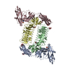
| Entry | Database: PDB / ID: 5xnp | ||||||
|---|---|---|---|---|---|---|---|
| Title | Crystal structures of human SALM5 in complex with human PTPdelta | ||||||
 Components Components |
| ||||||
 Keywords Keywords | MEMBRANE PROTEIN/HYDROLASE / Regulating some neural developments / MEMBRANE PROTEIN-HYDROLASE complex | ||||||
| Function / homology |  Function and homology information Function and homology informationtrans-synaptic signaling by trans-synaptic complex / cell surface receptor protein tyrosine phosphatase signaling pathway / negative regulation of macrophage activation / Receptor-type tyrosine-protein phosphatases / presynaptic membrane assembly / transmembrane receptor protein tyrosine phosphatase activity / synaptic membrane adhesion / presynapse assembly / regulation of postsynaptic density assembly / positive regulation of dendritic spine morphogenesis ...trans-synaptic signaling by trans-synaptic complex / cell surface receptor protein tyrosine phosphatase signaling pathway / negative regulation of macrophage activation / Receptor-type tyrosine-protein phosphatases / presynaptic membrane assembly / transmembrane receptor protein tyrosine phosphatase activity / synaptic membrane adhesion / presynapse assembly / regulation of postsynaptic density assembly / positive regulation of dendritic spine morphogenesis / Synaptic adhesion-like molecules / positive regulation of synapse assembly / phosphate-containing compound metabolic process / heterophilic cell-cell adhesion / regulation of presynapse assembly / protein-tyrosine-phosphatase / protein tyrosine phosphatase activity / cell adhesion molecule binding / hippocampal mossy fiber to CA3 synapse / negative regulation of inflammatory response / postsynaptic density membrane / modulation of chemical synaptic transmission / Schaffer collateral - CA1 synapse / GABA-ergic synapse / neuron differentiation / nervous system development / presynaptic membrane / signaling receptor binding / glutamatergic synapse / cell surface / signal transduction / extracellular exosome / plasma membrane Similarity search - Function | ||||||
| Biological species |  Homo sapiens (human) Homo sapiens (human) | ||||||
| Method |  X-RAY DIFFRACTION / SYNCHROTRON / MOLECULAR REPLACEMENT / Resolution: 3.729 Å X-RAY DIFFRACTION / SYNCHROTRON / MOLECULAR REPLACEMENT / Resolution: 3.729 Å | ||||||
 Authors Authors | Liu, H. / Lin, Z. / Xu, F. | ||||||
 Citation Citation | Journal: Nat Commun / Year: 2018 Title: Structural basis of SALM5-induced PTP delta dimerization for synaptic differentiation Authors: Lin, Z. / Liu, J. / Ding, H. / Xu, F. / Liu, H. | ||||||
| History |
|
- Structure visualization
| Structure viewer | Molecule: MolmilJmol/JSmol |
|---|
- Downloads & links
-Download
| PDBx/mmCIF format | 5xnp.cif.gz | 272.9 KB | Display | PDBx/mmCIF format |
|---|---|---|---|---|
| PDB format | pdb5xnp.ent.gz | 217.7 KB | Display | PDB format |
| PDBx/mmJSON format | 5xnp.json.gz | Tree view | PDBx/mmJSON format | |
| Others |  Other downloads Other downloads |
-Validation report
| Arichive directory | https://data.pdbj.org/pub/pdb/validation_reports/xn/5xnpftp://data.pdbj.org/pub/pdb/validation_reports/xn/5xnp | HTTPS FTP |
|---|
-Related structure data
| Related structure data |  5xnqSC 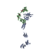 4yh7S S: Starting model for refinement C: citing same article ( |
|---|---|
| Similar structure data |


-Links
 PDBj
PDBj


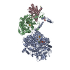
- Assembly
| Deposited unit | 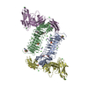
| ||||||||
|---|---|---|---|---|---|---|---|---|---|
| 1 |
| ||||||||
| Unit cell |
|
-Components
-Protein , 2 types, 4 molecules ABDE
| #1: Protein | Mass: 40468.383 Da / Num. of mol.: 2 / Fragment: UNP residues 18-374 Source method: isolated from a genetically manipulated source Source: (gene. exp.) Homo sapiens (human) / Gene: LRFN5, C14orf146, SALM5 / Production host: Homo sapiens (human) / References: UniProt: Q96NI6#2: Protein | Mass: 32046.260 Da / Num. of mol.: 2 / Fragment: UNP residues 21-320 / Mutation: 181-189 deletion Source method: isolated from a genetically manipulated source Source: (gene. exp.) Homo sapiens (human) / Gene: PTPRD / Production host: Homo sapiens (human) / References: UniProt: P23468, protein-tyrosine-phosphatase |
|---|
-Sugars , 1 types, 8 molecules 
| #3: Sugar | ChemComp-NAG /  Type: D-saccharide, beta linking / Mass: 221.208 Da / Num. of mol.: 8 Type: D-saccharide, beta linking / Mass: 221.208 Da / Num. of mol.: 8Source method: isolated from a genetically manipulated source Formula: C8H15NO6 |
|---|
-Non-polymers , 5 types, 124 molecules 


| #4: Chemical |  Mass: 22.990 Da / Num. of mol.: 2 / Source method: obtained synthetically / Formula: Na Mass: 22.990 Da / Num. of mol.: 2 / Source method: obtained synthetically / Formula: Na#5: Chemical | ChemComp-CA /  Mass: 40.078 Da / Num. of mol.: 12 / Source method: obtained synthetically / Formula: Ca Mass: 40.078 Da / Num. of mol.: 12 / Source method: obtained synthetically / Formula: Ca#6: Chemical | ChemComp-CL /  Mass: 35.453 Da / Num. of mol.: 12 / Source method: obtained synthetically / Formula: Cl Mass: 35.453 Da / Num. of mol.: 12 / Source method: obtained synthetically / Formula: Cl#7: Chemical | ChemComp-SO4 /  Mass: 96.063 Da / Num. of mol.: 4 / Source method: obtained synthetically / Formula: SO4 Mass: 96.063 Da / Num. of mol.: 4 / Source method: obtained synthetically / Formula: SO4#8: Water | ChemComp-HOH / | Mass: 18.015 Da / Num. of mol.: 94 / Source method: isolated from a natural source / Formula: H2O |
|---|
-Details
| Has protein modification | Y |
|---|
-Experimental details
-Experiment
| Experiment | Method: X-RAY DIFFRACTION / Number of used crystals: 1 |
|---|
- Sample preparation
| Crystal | Density Matthews: 4.46 Å3/Da / Density % sol: 72.4 % |
|---|---|
| Crystal grow | Temperature: 293 K / Method: vapor diffusion, sitting drop / pH: 6 / Details: Tartrate Na/K, MES pH 6.0, lithium sulfate |
-Data collection
| Diffraction | Mean temperature: 100 K |
|---|---|
| Diffraction source | Source: SYNCHROTRON / Site: SSRF  / Beamline: BL19U1 / Wavelength: 0.987 Å / Beamline: BL19U1 / Wavelength: 0.987 Å |
| Detector | Type: DECTRIS PILATUS3 S 6M / Detector: PIXEL / Date: Jan 18, 2017 |
| Radiation | Protocol: SINGLE WAVELENGTH / Monochromatic (M) / Laue (L): M / Scattering type: x-ray |
| Radiation wavelength | Wavelength: 0.987 Å / Relative weight: 1 |
| Reflection | Resolution: 3.729→50 Å / Num. obs: 26924 / % possible obs: 99.8 % / Redundancy: 7.1 % / Rmerge(I) obs: 0.109 / Χ2: 1.563 / Net I/σ(I): 18.75 |
| Reflection shell | Resolution: 3.729→3.79 Å / Redundancy: 7.2 % / Rmerge(I) obs: 0.919 / Mean I/σ(I) obs: 2.5 / Num. unique obs: 1340 / CC1/2: 0.727 / Χ2: 1.656 / % possible all: 99.9 |
- Processing
| Software |
| ||||||||||||||||||||||||||||||||||||||||||||||||||||||||||||||||||||||
|---|---|---|---|---|---|---|---|---|---|---|---|---|---|---|---|---|---|---|---|---|---|---|---|---|---|---|---|---|---|---|---|---|---|---|---|---|---|---|---|---|---|---|---|---|---|---|---|---|---|---|---|---|---|---|---|---|---|---|---|---|---|---|---|---|---|---|---|---|---|---|---|
| Refinement | Method to determine structure: MOLECULAR REPLACEMENT Starting model: 5XNQ, 4YH7 Resolution: 3.729→48.908 Å / SU ML: 0.43 / Cross valid method: FREE R-VALUE / σ(F): 1.35 / Phase error: 27.44
| ||||||||||||||||||||||||||||||||||||||||||||||||||||||||||||||||||||||
| Solvent computation | Shrinkage radii: 0.9 Å / VDW probe radii: 1.11 Å | ||||||||||||||||||||||||||||||||||||||||||||||||||||||||||||||||||||||
| Refinement step | Cycle: LAST / Resolution: 3.729→48.908 Å
| ||||||||||||||||||||||||||||||||||||||||||||||||||||||||||||||||||||||
| Refine LS restraints |
| ||||||||||||||||||||||||||||||||||||||||||||||||||||||||||||||||||||||
| LS refinement shell |
|
