Movie
Movie Controller

[English] 日本語
 Yorodumi
- PDB-5x5b: Prefusion structure of SARS-CoV spike glycoprotein, conformation 2 -
+ Open data

- Basic information
| Entry | Database: PDB / ID: 5x5b | ||||||||||||||||||||||||||||||||||||
|---|---|---|---|---|---|---|---|---|---|---|---|---|---|---|---|---|---|---|---|---|---|---|---|---|---|---|---|---|---|---|---|---|---|---|---|---|---|
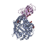
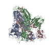
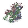
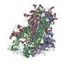
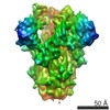
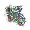
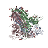
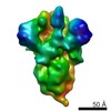
| Title | Prefusion structure of SARS-CoV spike glycoprotein, conformation 2 | ||||||||||||||||||||||||||||||||||||
 Components Components | Spike glycoprotein | ||||||||||||||||||||||||||||||||||||
 Keywords Keywords | VIRAL PROTEIN / SARS-CoV / spike glycoprotein / prefusion / single particle | ||||||||||||||||||||||||||||||||||||
| Function / homology |  Function and homology information Function and homology informationMaturation of spike protein / Translation of Structural Proteins / Virion Assembly and Release / Attachment and Entry / SARS-CoV-1 activates/modulates innate immune responses / symbiont-mediated-mediated suppression of host tetherin activity / membrane fusion / host cell endoplasmic reticulum-Golgi intermediate compartment membrane / positive regulation of viral entry into host cell / receptor-mediated virion attachment to host cell ...Maturation of spike protein / Translation of Structural Proteins / Virion Assembly and Release / Attachment and Entry / SARS-CoV-1 activates/modulates innate immune responses / symbiont-mediated-mediated suppression of host tetherin activity / membrane fusion / host cell endoplasmic reticulum-Golgi intermediate compartment membrane / positive regulation of viral entry into host cell / receptor-mediated virion attachment to host cell / host cell surface receptor binding / symbiont-mediated suppression of host innate immune response / endocytosis involved in viral entry into host cell / fusion of virus membrane with host plasma membrane / fusion of virus membrane with host endosome membrane / viral envelope / host cell plasma membrane / virion membrane / identical protein binding / membrane Similarity search - Function | ||||||||||||||||||||||||||||||||||||
| Biological species |  SARS coronavirus BJ01 SARS coronavirus BJ01 | ||||||||||||||||||||||||||||||||||||
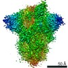
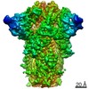
| Method | ELECTRON MICROSCOPY / single particle reconstruction / cryo EM / Resolution: 3.7 Å | ||||||||||||||||||||||||||||||||||||
 Authors Authors | Yuan, Y. / Cao, D. / Zhang, Y. / Ma, J. / Qi, J. / Wang, Q. / Lu, G. / Wu, Y. / Yan, J. / Shi, Y. ...Yuan, Y. / Cao, D. / Zhang, Y. / Ma, J. / Qi, J. / Wang, Q. / Lu, G. / Wu, Y. / Yan, J. / Shi, Y. / Zhang, X. / Gao, G.F. | ||||||||||||||||||||||||||||||||||||
| Funding support |  China, 11items China, 11items
| ||||||||||||||||||||||||||||||||||||
 Citation Citation | Journal: Nat Commun / Year: 2017 Title: Cryo-EM structures of MERS-CoV and SARS-CoV spike glycoproteins reveal the dynamic receptor binding domains. Authors: Yuan Yuan / Duanfang Cao / Yanfang Zhang / Jun Ma / Jianxun Qi / Qihui Wang / Guangwen Lu / Ying Wu / Jinghua Yan / Yi Shi / Xinzheng Zhang / George F Gao / Abstract: The envelope spike (S) proteins of MERS-CoV and SARS-CoV determine the virus host tropism and entry into host cells, and constitute a promising target for the development of prophylactics and ...The envelope spike (S) proteins of MERS-CoV and SARS-CoV determine the virus host tropism and entry into host cells, and constitute a promising target for the development of prophylactics and therapeutics. Here, we present high-resolution structures of the trimeric MERS-CoV and SARS-CoV S proteins in its pre-fusion conformation by single particle cryo-electron microscopy. The overall structures resemble that from other coronaviruses including HKU1, MHV and NL63 reported recently, with the exception of the receptor binding domain (RBD). We captured two states of the RBD with receptor binding region either buried (lying state) or exposed (standing state), demonstrating an inherently flexible RBD readily recognized by the receptor. Further sequence conservation analysis of six human-infecting coronaviruses revealed that the fusion peptide, HR1 region and the central helix are potential targets for eliciting broadly neutralizing antibodies. | ||||||||||||||||||||||||||||||||||||
| History |
|
- Structure visualization
| Movie |
Movie viewer |
|---|---|
| Structure viewer | Molecule: MolmilJmol/JSmol |
- Downloads & links
-Download
| PDBx/mmCIF format | 5x5b.cif.gz | 632.2 KB | Display | PDBx/mmCIF format |
|---|---|---|---|---|
| PDB format | pdb5x5b.ent.gz | 502.7 KB | Display | PDB format |
| PDBx/mmJSON format | 5x5b.json.gz | Tree view | PDBx/mmJSON format | |
| Others |  Other downloads Other downloads |
-Validation report
| Summary document | 5x5b_validation.pdf.gz | 832.2 KB | Display | wwPDB validaton report |
|---|---|---|---|---|
| Full document | 5x5b_full_validation.pdf.gz | 874.3 KB | Display | |
| Data in XML | 5x5b_validation.xml.gz | 87.3 KB | Display | |
| Data in CIF | 5x5b_validation.cif.gz | 131.9 KB | Display | |
| Arichive directory | https://data.pdbj.org/pub/pdb/validation_reports/x5/5x5bftp://data.pdbj.org/pub/pdb/validation_reports/x5/5x5b | HTTPS FTP |
-Related structure data
| Related structure data | 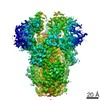 6705MC 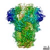 6703C 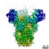 6704C 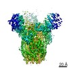 6706C 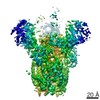 6707C 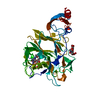 5x4rC 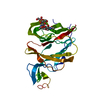 5x4sC 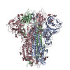 5x58C 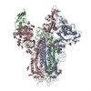 5x59C 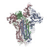 5x5cC 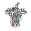 5x5fC C: citing same article ( M: map data used to model this data |
|---|---|
| Similar structure data |

-Links
 PDBj
PDBj

- Assembly
| Deposited unit | 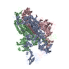
|
|---|---|
| 1 |
|
-Components
| #1: Protein | Mass: 136825.891 Da / Num. of mol.: 3 Source method: isolated from a genetically manipulated source Source: (gene. exp.) SARS coronavirus BJ01 / Strain: BJ01 / Production host:   Spodoptera frugiperda (fall armyworm) / References: UniProt: P59594*PLUS Spodoptera frugiperda (fall armyworm) / References: UniProt: P59594*PLUSHas protein modification | Y | Sequence details | THE REFERENCE SEQUENCE DATABASE OF THIS PROTEIN IS RESIDUES 14-1193 IN GENBANK AAP30030.1. RESIDUES ...THE REFERENCE SEQUENCE DATABASE OF THIS PROTEIN IS RESIDUES 14-1193 IN GENBANK AAP30030.1. RESIDUES 1194-1241 ARE EXPRESSION | |
|---|
-Experimental details
-Experiment
| Experiment | Method: ELECTRON MICROSCOPY |
|---|---|
| EM experiment | Aggregation state: PARTICLE / 3D reconstruction method: single particle reconstruction |
- Sample preparation
| Component | Name: SARS-CoV spike trimer / Type: COMPLEX / Entity ID: all / Source: RECOMBINANT |
|---|---|
| Source (natural) | Organism: SARS coronavirus BJ01 / Strain: BJ01 |
| Source (recombinant) | Organism: Spodoptera frugiperda (fall armyworm) |
| Buffer solution | pH: 7.5 |
| Specimen | Embedding applied: NO / Shadowing applied: NO / Staining applied: NO / Vitrification applied: YES |
| Vitrification | Cryogen name: ETHANE |
- Electron microscopy imaging
| Experimental equipment |  Model: Titan Krios / Image courtesy: FEI Company |
|---|---|
| Microscopy | Model: FEI TITAN KRIOS |
| Electron gun | Electron source:  FIELD EMISSION GUN / Accelerating voltage: 300 kV / Illumination mode: FLOOD BEAM FIELD EMISSION GUN / Accelerating voltage: 300 kV / Illumination mode: FLOOD BEAM |
| Electron lens | Mode: BRIGHT FIELD |
| Image recording | Electron dose: 8 e/Å2 / Film or detector model: GATAN K2 SUMMIT (4k x 4k) |
- Processing
| Software | Name: PHENIX / Version: dev_2411: / Classification: refinement | ||||||||||||||||||||||||
|---|---|---|---|---|---|---|---|---|---|---|---|---|---|---|---|---|---|---|---|---|---|---|---|---|---|
| CTF correction | Type: PHASE FLIPPING AND AMPLITUDE CORRECTION | ||||||||||||||||||||||||
| 3D reconstruction | Resolution: 3.7 Å / Resolution method: FSC 0.143 CUT-OFF / Num. of particles: 60000 / Symmetry type: POINT | ||||||||||||||||||||||||
| Atomic model building | Protocol: RIGID BODY FIT | ||||||||||||||||||||||||
| Atomic model building | 3D fitting-ID: 1 / Source name: PDB / Type: experimental model
| ||||||||||||||||||||||||
| Refine LS restraints |
|