 Movie
Movie Controller

[English] 日本語
 Yorodumi
- EMDB-6707: Prefusion structure of MERS-CoV spike glycoprotein, conformation 2 -
+ Open data

- Basic information
| Entry | Database: EMDB / ID: EMD-6707 | |||||||||
|---|---|---|---|---|---|---|---|---|---|---|
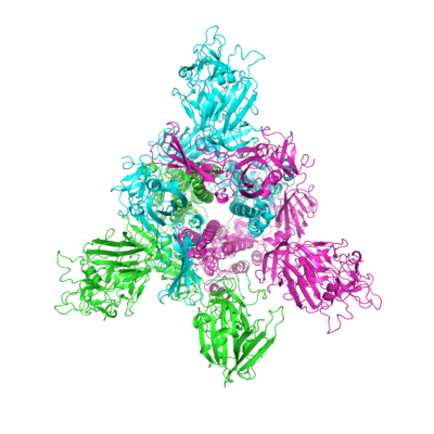
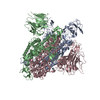
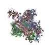
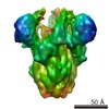
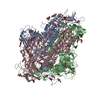
| Title | Prefusion structure of MERS-CoV spike glycoprotein, conformation 2 | |||||||||
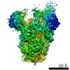
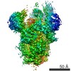
 Map data Map data | ||||||||||
 Sample Sample |
| |||||||||
 Keywords Keywords | MERS-CoV / spike glycoprotein / prefusion / single particle / VIRAL PROTEIN | |||||||||
| Function / homology |  Function and homology information Function and homology informationpositive regulation of viral entry into host cell / membrane fusion / host cell endoplasmic reticulum-Golgi intermediate compartment membrane / receptor-mediated virion attachment to host cell / endocytosis involved in viral entry into host cell / fusion of virus membrane with host plasma membrane / fusion of virus membrane with host endosome membrane / viral envelope / host cell plasma membrane / virion membrane / membrane Similarity search - Function | |||||||||
| Biological species |  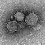 Middle East respiratory syndrome coronavirus Middle East respiratory syndrome coronavirus | |||||||||
| Method | single particle reconstruction / cryo EM / Resolution: 4.2 Å | |||||||||
 Authors Authors | Yuan Y / Cao D | |||||||||
 Citation Citation | Journal: Nat Commun / Year: 2017 Title: Cryo-EM structures of MERS-CoV and SARS-CoV spike glycoproteins reveal the dynamic receptor binding domains. Authors: Yuan Yuan / Duanfang Cao / Yanfang Zhang / Jun Ma / Jianxun Qi / Qihui Wang / Guangwen Lu / Ying Wu / Jinghua Yan / Yi Shi / Xinzheng Zhang / George F Gao /  Abstract: The envelope spike (S) proteins of MERS-CoV and SARS-CoV determine the virus host tropism and entry into host cells, and constitute a promising target for the development of prophylactics and ...The envelope spike (S) proteins of MERS-CoV and SARS-CoV determine the virus host tropism and entry into host cells, and constitute a promising target for the development of prophylactics and therapeutics. Here, we present high-resolution structures of the trimeric MERS-CoV and SARS-CoV S proteins in its pre-fusion conformation by single particle cryo-electron microscopy. The overall structures resemble that from other coronaviruses including HKU1, MHV and NL63 reported recently, with the exception of the receptor binding domain (RBD). We captured two states of the RBD with receptor binding region either buried (lying state) or exposed (standing state), demonstrating an inherently flexible RBD readily recognized by the receptor. Further sequence conservation analysis of six human-infecting coronaviruses revealed that the fusion peptide, HR1 region and the central helix are potential targets for eliciting broadly neutralizing antibodies. | |||||||||
| History |
|
- Structure visualization
| Movie |
Movie viewer |
|---|---|
| Structure viewer | EM map: SurfViewMolmilJmol/JSmol |
| Supplemental images |
- Downloads & links
-EMDB archive
| Map data | emd_6707.map.gz | 28.4 MB | EMDB map data format | |
|---|---|---|---|---|
| Header (meta data) | emd-6707-v30.xmlemd-6707.xml | 9.9 KB 9.9 KB | Display Display | EMDB header |
| Images |  emd_6707.png emd_6707.png | 120.4 KB | ||
| Filedesc metadata | emd-6707.cif.gz | 5.6 KB | ||
| Archive directory |  http://ftp.pdbj.org/pub/emdb/structures/EMD-6707ftp://ftp.pdbj.org/pub/emdb/structures/EMD-6707 http://ftp.pdbj.org/pub/emdb/structures/EMD-6707ftp://ftp.pdbj.org/pub/emdb/structures/EMD-6707 | HTTPS FTP |
-Related structure data
| Related structure data |  5x5fMC 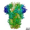 6703C 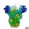 6704C 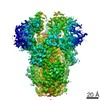 6705C 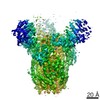 6706C 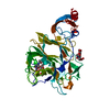 5x4rC 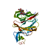 5x4sC 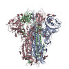 5x58C 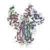 5x59C 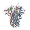 5x5bC 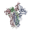 5x5cC M: atomic model generated by this map C: citing same article ( |
|---|---|
| Similar structure data |
-Links
| EMDB pages | EMDB (EBI/PDBe) / EMDataResource |
|---|
-Map
| File | Download / File: emd_6707.map.gz / Format: CCP4 / Size: 30.5 MB / Type: IMAGE STORED AS FLOATING POINT NUMBER (4 BYTES) | ||||||||||||||||||||||||||||||||||||||||||||||||||||||||||||
|---|---|---|---|---|---|---|---|---|---|---|---|---|---|---|---|---|---|---|---|---|---|---|---|---|---|---|---|---|---|---|---|---|---|---|---|---|---|---|---|---|---|---|---|---|---|---|---|---|---|---|---|---|---|---|---|---|---|---|---|---|---|
| Projections & slices | Image control
Images are generated by Spider. | ||||||||||||||||||||||||||||||||||||||||||||||||||||||||||||
| Voxel size | X=Y=Z: 1.3 Å | ||||||||||||||||||||||||||||||||||||||||||||||||||||||||||||
| Density |
| ||||||||||||||||||||||||||||||||||||||||||||||||||||||||||||
| Symmetry | Space group: 1 | ||||||||||||||||||||||||||||||||||||||||||||||||||||||||||||
| Details | EMDB XML:
CCP4 map header:
| ||||||||||||||||||||||||||||||||||||||||||||||||||||||||||||
 Z (Sec.)
 Y (Row.)
 X (Col.)


-Supplemental data
- Sample components
-Entire : MERS-CoV spike trimer
| Entire | Name: MERS-CoV spike trimer |
|---|---|
| Components |
|
-Supramolecule #1: MERS-CoV spike trimer
| Supramolecule | Name: MERS-CoV spike trimer / type: complex / ID: 1 / Parent: 0 / Macromolecule list: all |
|---|---|
| Source (natural) | Organism: Middle East respiratory syndrome coronavirus |
-Macromolecule #1: S protein
| Macromolecule | Name: S protein / type: protein_or_peptide / ID: 1 / Number of copies: 3 / Enantiomer: LEVO |
|---|---|
| Source (natural) | Organism: Middle East respiratory syndrome coronavirus |
| Molecular weight | Theoretical: 145.856203 KDa |
| Recombinant expression | Organism:   Spodoptera frugiperda (fall armyworm) Spodoptera frugiperda (fall armyworm) |
| Sequence | String: YVDVGPDSVK SACIEVDIQQ TFFDKTWPRP IDVSKADGII YPQGRTYSNI TITYQGLFPY QGDHGDMYVY SAGHATGTTP QKLFVANYS QDVKQFANGF VVRIGAAANS TGTVIISPST SATIRKIYPA FMLGSSVGNF SDGKMGRFFN HTLVLLPDGC G TLLRAFYC ...String: YVDVGPDSVK SACIEVDIQQ TFFDKTWPRP IDVSKADGII YPQGRTYSNI TITYQGLFPY QGDHGDMYVY SAGHATGTTP QKLFVANYS QDVKQFANGF VVRIGAAANS TGTVIISPST SATIRKIYPA FMLGSSVGNF SDGKMGRFFN HTLVLLPDGC G TLLRAFYC ILEPRSGNHC PAGNSYTSFA TYHTPATDCS DGNYNRNASL NSFKEYFNLR NCTFMYTYNI TEDEILEWFG IT QTAQGVH LFSSRYVDLY GGNMFQFATL PVYDTIKYYS IIPHSIRSIQ SDRKAWAAFY VYKLQPLTFL LDFSVDGYIR RAI DCGFND LSQLHCSYES FDVESGVYSV SSFEAKPSGS VVEQAEGVEC DFSPLLSGTP PQVYNFKRLV FTNCNYNLTK LLSL FSVND FTCSQISPAA IASNCYSSLI LDYFSYPLSM KSDLSVSSAG PISQFNYKQS FSNPTCLILA TVPHNLTTIT KPLKY SYIN KCSRLLSDDR TEVPQLVNAN QYSPCVSIVP STVWEDGDYY RKQLSPLEGG GWLVASGSTV AMTEQLQMGF GITVQY GTD TNSVCPKLEF ANDTKIASQL GNCVEYSLYG VSGRGVFQNC TAVGVRQQRF VYDAYQNLVG YYSDDGNYYC LRACVSV PV SVIYDKETKT HATLFGSVAC EHISSTMSQY SRSTRSMLKR RDSTYGPLQT PVGCVLGLVN SSLFVEDCKL PLGQSLCA L PDTPSTLTPR SVSSVPGEMR LASIAFNHPI QVDQLNSSYF KLSIPTNFSF GVTQEYIQTT IQKVTVDCKQ YVCNGFQKC EQLLREYGQF CSKINQALHG ANLRQDDSVR NLFASVKSSQ SSPIIPGFGG DFNLTLLEPV SISTGSRSAR SAIEDLLFDK VTIADPGYM QGYDDCMQQG PASARDLICA QYVAGYKVLP PLMDVNMEAA YTSSLLGSIA GVGWTAGLSS FAAIPFAQSI F YRLNGVGI TQQVLSENQK LIANKFNQAL GAMQTGFTTT NEAFQKVQDA VNNNAQALSK LASELSNTFG AISASIGDII QR LDVLEQD AQIDRLINGR LTTLNAFVAQ QLVRSESAAL SAQLAKDKVN ECVKAQSKRS GFCGQGTHIV SFVVNAPNGL YFM HVGYYP SNHIEVVSAY GLCDAANPTN CIAPVNGYFI KTNNTRIVDE WSYTGSSFYA PEPITSLNTK YVAPQVTYQN ISTN LPPPL LGNSTGIDFQ DELDEFFKNV STSIPNFGSL TQINTTLLDL TYEMLSLQQV VKALNESYID LKELGNYTYY NKEFR LVPR GSPGSGYIPE APRDGQAYVR KDGEWVLLST FLGHHHHHH UniProtKB: Spike glycoprotein |
-Experimental details
-Structure determination
| Method | cryo EM |
|---|---|
 Processing Processing | single particle reconstruction |
| Aggregation state | particle |
-Sample preparation
| Buffer | pH: 7.5 |
|---|---|
| Vitrification | Cryogen name: ETHANE |
- Electron microscopy
| Microscope | FEI TITAN KRIOS |
|---|---|
| Image recording | Film or detector model: GATAN K2 SUMMIT (4k x 4k) / Average electron dose: 8.0 e/Å2 |
| Electron beam | Acceleration voltage: 300 kV / Electron source:  FIELD EMISSION GUN FIELD EMISSION GUN |
| Electron optics | Illumination mode: FLOOD BEAM / Imaging mode: BRIGHT FIELD |
| Experimental equipment |  Model: Titan Krios / Image courtesy: FEI Company |
-Image processing
| Startup model | Type of model: EMDB MAP |
|---|---|
| Final reconstruction | Resolution.type: BY AUTHOR / Resolution: 4.2 Å / Resolution method: FSC 0.143 CUT-OFF / Number images used: 60000 |
| Initial angle assignment | Type: PROJECTION MATCHING |
| Final angle assignment | Type: PROJECTION MATCHING |
